Visual acuity in patients with retinitis pigmentosa
Abstract
Objective: The present study is taken up to learn the visual acuity in patients with retinitis pigmentosa (RP).
Methods: The current study was conducted in thirty male and female patients with in age group of 30-50 years and thirty age matched controls with retinitis pigmentosa. Vision testing was conducted by trained optometrician. Visual acuity was tested for distance vision using Snellen's test type placed at a distance of 6 meters from the student and near vision using near vision test type with the student holding the chart in his/her hand at a distance of approximately 30 cms from the face. Data was analyzed by SPSS 20.0. Statistical test used are student t test. P value less than 0.05 was considered significant.
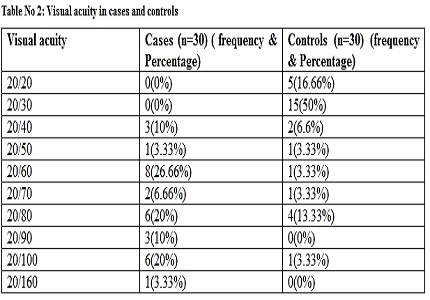
Results: There was no significant difference between cases and controls in demographic data. Decline in the visual acuity was more prevalent in cases when compared to controls.
Conclusion: We have observed decline in the visual acuity in the patients with retinitis pigmentosa. We recommend further detailed studies in this area.
Downloads
References
2. Regan D, Neima D. Low-contrast letter charts as a test of visual function. Ophthalmology. 1983 Oct;90(10):1192-200. [PubMed]
3. Maho Oishi, Hajime Nakamura, Masanori Hangai, Akio Oishi, Atsushi Otani, and Nagahisa Yoshimura. Contrast visual acuity in patients with retinitis pigmentosa assessed by a contrast sensitivity tester. Indian J Ophthalmol. 2012; 60(6): 545–549.
4. Alexander KR, Derlacki DJ, Fishman GA. Visual acuity vs letter contrast sensitivity in retinitis pigmentosa. Vision Res. 1995 May;35(10):1495-9. [PubMed]
5. Oomachi K, Ogata K, Sugawara T, Hagiwara A, Hata A, Yamamoto S. Evaluation of contrast visual acuity in patients with retinitis pigmentosa. Clin Ophthalmol. 2011;5:1459-63. doi: 10.2147/OPTH.S23070. Epub 2011 Oct 11. [PubMed]
6. Spellman DC, Alexander KR, Fishman GA, Derlacki DJ. Letter contrast sensitivity in retinitis pigmentosa patients assessed by Regan charts. Retina. 1989;9(4):287-91.
7. Alexander KR, Derlacki DJ, Fishman GA. Contrast thresholds for letter identification in retinitis pigmentosa. Invest Ophthalmol Vis Sci. 1992 May;33(6):1846-52.
8. A Datta, N Bhardwaj, SR Patrikar,and R Bhalwar. Study of Disorders of Visual Acuity among Adolescent School Children in Pune. Med J Armed Forces India. 2009; 65(1): 26–29. [PubMed]
9. Bunker CH, Berson EL, Bromley WC, Hayes RP, Roderick TH. Prevalence of retinitis pigmentosa in Maine. Am J Ophthalmol. 1984 Mar;97(3):357-65. [PubMed]
10. Novak-Lauš K, Suzana Kukulj S, Zoric-Geber M, Bastaic O. Primary tapetoretinal dystrophies as the cause of blindness and impaired vision in the republic of Croatia. Acta Clin Croat. 2002; 41(1): 23–27.
11. Grøndahl J. Estimation of prognosis and prevalence of retinitis pigmentosa and Usher syndrome in Norway. Clin Genet. 1987 Apr;31(4):255-64. [PubMed]
12. Hata H, Yonezawa M, Nakanishi T, Ri T, Yanashima K. Causes of entering institutions for visually handicapped persons during the past fi fteen years. Jpn J Clin Ophthalmol. 2003; 57: 259–62.
13. Buch H, Vinding T, La Cour M, Appleyard M, Jensen GB, Nielsen NV. Prevalence and causes of visual impairment and blindness among 9980 Scandinavian adults: the Copenhagen City Eye Study. Ophthalmology. 2004 Jan;111(1):53-61.
14. Bunker CH, Berson EL, Bromley WC, Hayes RP, Roderick TH. Prevalence of retinitis pigmentosa in Maine. Am J Ophthalmol. 1984 Mar;97(3):357-65. [PubMed]
15. Grøndahl J. Estimation of prognosis and prevalence of retinitis pigmentosa and Usher syndrome in Norway. Clin Genet. 1987 Apr;31(4):255-64. [PubMed]
16. Novak-Lauš K, Suzana Kukulj S, Zoric-Geber M, Bastaic O. Primary tapetoretinal dystrophies as the cause of blindness and impaired vision in the republic of Croatia. Acta Clin Croat 2002; 41: 23–27.
17. Al-Merjan JI, Pandova MG, Al-Ghanim M, Al-Wayel A, Al-Mutairi S. Registered blindness and low vision in Kuwait. Ophthalmic Epidemiol. 2005 Aug;12(4):251-7. [PubMed]
18. Buch H, Vinding T, La Cour M, Appleyard M, Jensen GB, Nielsen NV. Prevalence and causes of visual impairment and blindness among 9980 Scandinavian adults: the Copenhagen City Eye Study. Ophthalmology. 2004 Jan;111(1):53-61.
19. Hata H, Yonezawa M, Nakanishi T, Ri T, Yanashima K. Causes of entering institutions for visually handicapped persons during the past fi fteen years. Jpn J Clin Ophthalmol 2003; 57: 259–62.
20. Pennings RJ, Fields RR, Huygen PL, Deutman AF, Kimberling WJ, Cremers CW. Usher syndrome type III can mimic other types of Usher syndrome. Ann Otol Rhinol Laryngol 2003; 112: 525–30.
21. Tieder M, Levy M, Gubler MC, Gagnadoux MF, Broyer M. Renal abnormalities in the Bardet-Biedl syndrome. Int J Pediatr Nephrol. 1982 Sep;3(3):199-203. [PubMed]
22. Beales PL, Elcioglu N, Woolf AS, Parker D, Flinter FA. New criteria for improved diagnosis of Bardet-Biedl syndrome: results of a population survey. J Med Genet 1999; 36: 437–46.
23. Birch DG, Anderson JL, Fish GE. Yearly rates of rod and cone functional loss in retinitis pigmentosa and cone-rod dystrophy. Ophthalmology. 1999 Feb;106(2):258-68. [PubMed]
24. Grover S, Fishman GA, Anderson RJ, Tozatti MS, Heckenlively JR, Weleber RG, Edwards AO, Brown J Jr. Visual acuity impairment in patients with retinitis pigmentosa at age 45 years or older. Ophthalmology. 1999 Sep;106(9):1780-5. [PubMed]
25. Virgili G, Pierrottet C, Parmeggiani F, et al. Reading performance in patients with retinitis pigmentosa: a study using the MNREAD charts. Invest Ophthalmol Vis Sci 2004; 45: 3418–24.

Copyright (c) 2017 Author (s). Published by Siddharth Health Research and Social Welfare Society

This work is licensed under a Creative Commons Attribution 4.0 International License.


 OAI - Open Archives Initiative


 Therapoid

