Brown’y points for guessing!- A case report of an unusual pigmented corneal plaque.
Abstract
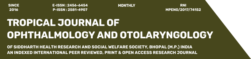
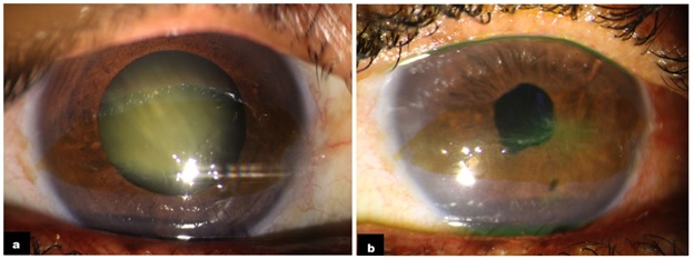
A 54-year-old female presented with complaints of blurring in the right eye since 2 years. A grade 3 nuclear sclerosis and a peculiar band-shaped subepithelial brown corneal patch, within the inter-palpebral area was noted. The overlying epithelium was scraped for smears and cultures which came back negative. The patient underwent uneventful cataract surgery, but returned 2 weeks later with complaints of watering and grittiness. Surprisingly a dendritic ulcer was noted within the pigmented patch, which responded to topical antivirals and tear substitutes. History of a similar episode, 3 years back was elicited upon questioning the patient.
Downloads
References
Darougar S, Wishart MS, Viswalingam ND. Epidemiological and clinical features of primary herpes simplex virus ocular infection. Br J Ophthalmol. 1985 Jan;69(1):2-6. doi: 10.1136/bjo.69.1.2.
Holland EJ, Schwartz GS. Classification of herpes simplex virus keratitis. Cornea. 1999 Mar;18(2):144-54. doi: 10.1097/00003226-199903000-00002.
Toma HS, Murina AT, Areaux RG Jr, Neumann DM, Bhattacharjee PS, Foster TP, Kaufman HE, Hill JM. Ocular HSV-1 latency, reactivation and recurrent disease. Semin Ophthalmol. 2008 Jul-Aug;23(4):249-73. doi: 10.1080/08820530802111085.
Perng GC, Jones C. Towards an understanding of the herpes simplex virus type 1 latency-reactivation cycle. Interdiscip Perspect Infect Dis. 2010;2010:262415. doi: 10.1155/2010/262415.
Shimmura S, Suematsu M, Shimoyama M, Tsubota K, Oguchi Y, Ishimura Y. Subthreshold UV radiation-induced peroxide formation in cultured corneal epithelial cells: the protective effects of lactoferrin. Exp Eye Res. 1996 Nov;63(5):519-26. doi: 10.1006/exer.1996.0142.
Ellison RT 3rd. The effects of lactoferrin on gram-negative bacteria. Adv Exp Med Biol. 1994;357:71-90. doi: 10.1007/978-1-4615-2548-6_8.
Loh A, Hadziahmetovic M, Dunaief JL. Iron homeostasis and eye disease. Biochim Biophys Acta. 2009 Jul;1790(7):637-49. doi: 10.1016/j.bbagen.2008.11.001.
Assil KK, Quantock AJ, Barrett AM, Schanzlin DJ. Corneal iron lines associated with the intrastromal corneal ring. Am J Ophthalmol. 1993 Sep 15;116(3):350-6. doi: 10.1016/s0002-9394(14)71353-4.
Hiratsuka Y, Nakayasu K, Kanai A. Secondary keratoconus with corneal epithelial iron ring similar to Fleischer's ring. Jpn J Ophthalmol. 2000 Jul-Aug;44(4):381-6. doi: 10.1016/s0021-5155(00)00179-9.
Copyright (c) 2021 Author (s). Published by Siddharth Health Research and Social Welfare Society

This work is licensed under a Creative Commons Attribution 4.0 International License.

 OAI - Open Archives Initiative


