Correlation between type 2 diabetes mellitus and central corneal thickness: A cross-sectional study
Abstract
Background and Aim: Diabetes has emerged as an important global health concern because of its various adverse effects on the ocular tissue. The present study was done to study the correlation between type 2 diabetes mellitus and central corneal thickness in patients coming to the tertiary care institute of Gujarat, India.
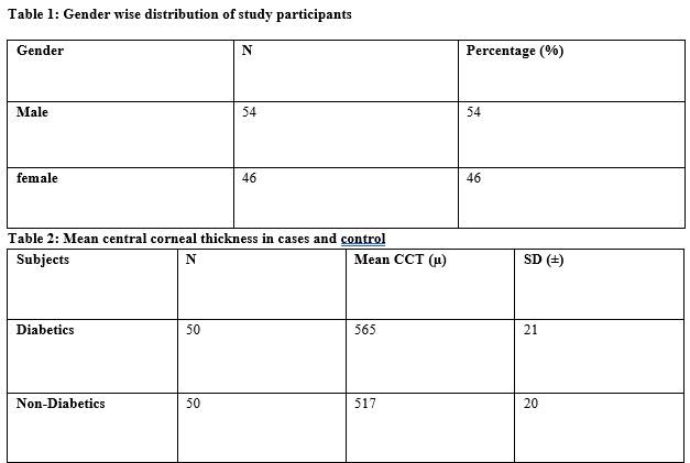
Material and Methods: The present study was conducted over 1 year at the tertiary care institute of Gujarat, India.50 patients with type 2 diabetes mellitus previously diagnosed by a physician on treatment and 50 age-matched controls who are non-diabetics on history and blood sugar levels were enrolled. The central corneal thickness was measured using an ultrasound pachymeter using multiple reading single point modes by a single person.
Results: The mean central corneal thickness in diabetics was 565 ± 21 micrometres and in non-diabetics was 517 ± 20 micrometres. The central corneal thickness was found to be higher in patients with type 2 diabetes mellitus when compared to non-diabetics.
Conclusion: Patients with type 2 diabetes mellitus were found to have thicker corneas as compared to non-diabetics. This should take into consideration while interpreting intraocular pressure and before any refractive surgeries in diabetics.
Downloads
References
Su DH, Wong TY, Wong WL, Saw SM, Tan DT, Shen SY, et al. Diabetes, hyperglycemia, and central corneal thickness: the Singapore Malay Eye Study. Ophthalmology. 2008 Jun;115(6):964-968.e1. doi: 10.1016/j.ophtha.2007.08.021.
Ozdamar Y, Cankaya B, Ozalp S, Acaroglu G, Karakaya J, Ozkan SS. Is there a correlation between diabetes mellitus and central corneal thickness? J Glaucoma. 2010 Dec;19(9):613-6. doi: 10.1097/IJG.0b013e3181ca7c62.
Schwartz DE. Corneal sensitivity in diabetics. Arch Ophthalmol. 1974 Mar;91(3):174-8. doi: 10.1001/archopht.1974.03900060182003.
Gekka M, Miyata K, Nagai Y, Nemoto S, Sameshima T, Tanabe T, et al. Corneal epithelial barrier function in diabetic patients. Cornea. 2004 Jan;23(1):35-7. doi: 10.1097/00003226-200401000-00006.
Naumann G.O.H, Holbach L, Kruse F.E. Apliedpathologyfor ophthalmic microsurgeons.Springer-verlag Berlin Heidelberg NewYork. 2008, Germany, 351.
Sahin A, Bayer A, Ozge G, Mumcuoğlu T. Corneal biomechanical changes in diabetes mellitus and their influence on intraocular pressure measurements. Invest Ophthalmol Vis Sci. 2009 Oct;50(10):4597-604. doi: 10.1167/iovs.08-2763. Epub 2009 May 14.
Schwartz DE. Corneal sensitivity in diabetics. Arch Ophthalmol. 1974 Mar;91(3):174-8. doi: 10.1001/archopht.1974.03900060182003.
Gekka M, Miyata K, Nagai Y, Nemoto S, Sameshima T, Tanabe T, et al. Corneal epithelial barrier function in diabetic patients. Cornea. 2004 Jan;23(1):35-7. doi: 10.1097/00003226-200401000-00006.
Roszkowska AM, Tringali CG, Colosi P, Squeri CA, Ferreri G. Corneal endothelium evaluation in type I and type II diabetes mellitus. Ophthalmologica. 1999;213(4):258-61. doi: 10.1159/000027431.
Keoleian GM, Pach JM, Hodge DO, Trocme SD, Bourne WM. Structural and functional studies of the corneal endothelium in diabetes mellitus. Am J Ophthalmol. 1992 Jan 15;113(1):64-70. doi: 10.1016/s0002-9394(14)757551.
Yaylali V, Kaufman SC, Thompson HW. Corneal thickness measurements with the Orbscan Topography System and ultrasonic pachymetry. J Cataract Refract Surg. 1997 Nov;23(9):1345-50. doi: 10.1016/s0886-3350(97)80113-7.
Salz JJ, Azen SP, Berstein J, Caroline P, Villasenor RA, Schanzlin DJ. Evaluation and comparison of sources of variability in the measurement of corneal thickness with ultrasonic and optical pachymeters. Ophthalmic Surg. 1983 Sep;14(9):750-4.
Williams R, Airey M, Baxter H, Forrester J, Kennedy-Martin T, Girach A. Epidemiology of diabetic retinopathy and macular oedema: a systematic review. Eye (Lond). 2004 Oct;18(10):963-83. doi: 10.1038/sj.eye.6701476.
Claramonte PJ, Ruiz-Moreno JM, Sánchez-Pérez SI, León M, Griñó C, Cerviño VD, et al. Variation of central corneal thickness in diabetic patients as detected by ultrasonic pachymetry. Arch Soc Esp Oftalmol. 2006 Sep;81(9):523-6. Spanish. doi: 10.4321/s0365-66912006000900007.
O'Donnell C, Efron N. Corneal endothelial cell morphometry and corneal thickness in diabetic contact lens wearers. Optom Vis Sci. 2004 Nov;81(11):858-62. doi: 10.1097/01.opx.0000145029.76675.f7.
Lee JS, Oum BS, Choi HY, Lee JE, Cho BM. Differences in corneal thickness and corneal endothelium related to duration in diabetes. Eye (Lond). 2006 Mar;20(3):315-8. doi: 10.1038/sj.eye.6701868.
Keoleian GM, Pach JM, Hodge DO, Trocme SD, Bourne WM. Structural and functional studies of the corneal endothelium in diabetes mellitus. Am J Ophthalmol. 1992 Jan 15;113(1):64-70. doi: 10.1016/s0002-9394(14)757551.
Ozdamar Y, Cankaya B, Ozalp S, Acaroglu G, Karakaya J, Ozkan SS. Is there a correlation between diabetes mellitus and central corneal thickness? J Glaucoma. 2010 Dec;19(9):613-6. doi: 10.1097/IJG.0b013e3181ca7c62.
Claramonte PJ, Ruiz-Moreno JM, Sánchez-Pérez SI, León M, Griñó C, Cerviño VD, et al. Variation of central corneal thickness in diabetic patients as detected by ultrasonic pachymetry. Arch Soc Esp Oftalmol. 2006 Sep;81(9):523-6. Spanish. doi: 10.4321/s0365-66912006000900007.
Lee JS, Oum BS, Choi HY, Lee JE, Cho BM. Differences in corneal thickness and corneal endothelium related to duration in diabetes. Eye (Lond). 2006 Mar;20(3):315-8. doi: 10.1038/sj.eye.6701868.
McNamara NA, Brand RJ, Polse KA, Bourne WM. Corneal function during normal and high serum glucose levels in diabetes. Invest Ophthalmol Vis Sci. 1998 Jan;39(1):3-17.
Yazgan S, Celik U, Kaldırım H, Ayar O, Elbay A, Aykut V, et al. Evaluation of the relationship between corneal biomechanic and HbA1C levels in type 2 diabetes patients. Clin Ophthalmol. 2014 Aug 19;8:1549-53. doi: 10.2147/OPTH.S67984.

Copyright (c) 2021 Author (s). Published by Siddharth Health Research and Social Welfare Society

This work is licensed under a Creative Commons Attribution 4.0 International License.


 OAI - Open Archives Initiative


 Therapoid

