Congenital sensorineural hearing loss- A radiological (CT and MRI) study
Abstract
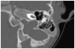
Aims: Congenital deafness refers to hearing loss which is believed to have been present since birth. The purpose of this study is to evaluate the clinical application of computed tomography (CT) and magnetic resonance imaging (MRI) in children with profound congenital deafness and to analyze anatomic abnormalities of the inner ear and the vestibulocochlear nerve.
Methods: A cross-sectional and observational study was carried out in Department of ENT of a tertiary care centre, in 50 cases of congenital deafness. All cases were analyzed for detailed history and underwent clinical audiological evaluations, high resolution computed tomography (CT) and 3D magnetic resonance imaging (MRI) of bilateral temporal bones.
Result: Out of total 50 patients, majority of them presented between 6 to 30 months of age. CT scan findings were found radiologically abnormal in 54% cases. Most commonly observed abnormality (21.82% in bilateral side) in CT scan was of middle ear cleft followed by vestibule (20% in bilateral side). MRI findings were found abnormal in 88% cases. Most commonly observed abnormality (39.51% in right side and 45.07% in left side) in MRI was of semi-circular canal followed by vestibules (16.05% in right side and 18.31% in left side).
Conclusion: All children with congenital deafness should undergo radiological investigation of the temporal bone and inner ear. The decision whether to perform a CT or MRI will depend on scanner availability and management considerations, but cochlear implant candidates will require both.
Downloads
References
2. Jackler RK, Luxford WM, House WF. Congenital Malformations of the inner ear: a classification based on embryogenesis. Laryngoscope; 1987; Mar; 97(3 Pt 2 Suppl 40):2-14.
3. Gleeson TG, Lacy PD, Bresnihan M, Gaffney R, Brennan P, Viani L. High resolution computed tomography and magnetic resonance imaging the pre-operative assessment of cochlear implant patients. J LaryngolOtol; 2003; 117 (9):692-95.doi: 10.1258/00222 1503322334495.
4. Lo WWM. Imaging of Cochlear and Auditory Brain Stem Implantation. Am J Neuroradiol; 1998; June-July; 19 (6): 1147-54.
5. Abdullah A, Mahmud MR, Maimunah A, Zulfiqar MA, Saim L, Mazlan R. Preoperative High Resolution CT and MR Imaging in Cochlear Implantation. Ann Acad Med Singapore; 2004; July; 32(4):442-5
6. Bath AP, O’DonoghueGM, Holland IM, Gibbin KP. Paediatric cochlear implantation: how reliable is computed tomography in assessing cochlear patency? Clinical Otolaryngol Allied Sciences; 1993; Dec;18(6): 475-9.
7. Woolford TJ, Roberts GR, Hartley C, Ramsden RT. Etiology of hearing loss and cochlear computed tomography: findings in preimplant assessment. Ann Otol Rhinol Laryngol Suppl. 1995; Sep; 166:201-6.
8. BamiouDE, Phelps P, Sirimanna T. Temporal bone computed tomography findings inbilateral sensorineural hearing loss. Arch Dis Child; 2000; Mar; 82(3): 257–260.
9. Shusterman D, Handler SD, Marsh RR, Bilaniuk L, Tom LWC. Usefulness of computed tomographic scan in the evaluation of sensorineural hearing loss in children. Arch Otolaryngol Head Neck Surg; 1992; May; 118(5):501–3.
10. Kalsotra P, Kumar S, Gosh P, Mishra NK, Verma IC. A Study of Congenital and Early Acquired Impairment of Hearing. JK Science: Journal of Medical Education & Research; 2002; July-Sept; 4 (3), 136-143.
11. Westerhof JP, Rademaker J, Weber BP, Becker H. Congenital malformations of the inner ear and the vestibulocochlear nerve in children with sensorineural hearing loss: evaluation with CT and MRI. J Comput Assist Tomogr; 2001; Sep-Oct;25(5):719-26.
12. Lam WW, Hui Y, Au DK, Chow LC, Chan FL, Yu L, Wei WI. Radiological study of temporal bone in children with profound deafness before cochlear implant: CT vs magnetic resonance imaging. Zhonghua Er Bi Yan Hou Ke Za Zhi, 2002; Dec;37 (6): 440-2.
13. Parry DA, Booth T, Roland PS. Advantages of magnetic resonance imaging over computed tomography in preoperative evaluation of pediatric cochlear implant candidates. OtolNeurotol; 2005; Sep; 26 (5): 976-82.
Copyright (c) 2018 Author (s). Published by Siddharth Health Research and Social Welfare Society

This work is licensed under a Creative Commons Attribution 4.0 International License.

 OAI - Open Archives Initiative


