
A case report of oculofacial rosacea complicating to corneal infiltrates and vascularisation
Abstract
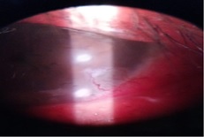
Purpose: To report a case of ocular rosacea with corneal infiltrates and vascularisation. Here we report a case of a 34-year-old male presenting with ocular rosacea with corneal infiltrates and vascularisation in the left eye with posterior blepharitis of both eyes. The clinical diagnosis was based on the facial findings of erythematous papulomacular lesions and telangiectasia. The patient was treated with systemic doxycycline and for ocular lesions treated with antibiotic steroids and lubricants. Ocular rosacea, if not treated on time, can worsen to the stage of corneal infiltrates and vascularisation.
Downloads
References
Audrey SC, Kathryn AC. Lid Inflammations. In: Albert DM, Jakobiec FA, Miller WJ (Eds).Principles and Practice of Ophthalmology:3rd ed. Philadelphia: Elsevier; 2008. pp 630-634.
Bowling B. Kanski’s Clinical Ophthalmology: A Systematic Approach. 8th ed. Edinburgh: Butterworth-Heinemann/Elsevier;2007.
Wilkin J, Dahl M, Detmar M, Drake L, Feinstein A, Odom R. et al. Standard classification of rosacea: Report of the National Rosacea Society expert committee on the classification and staging of rosacea. J Am Acad Dermatol. 2002;46(4):584–587. doi: https://doi.org/10.1067/mjd.2002.120625.
Akpek EK, Merchant A, Pinar V, Foster CS. Ocular rosacea: Patient characteristics and follow-up. Ophthalmol. 1997;104(11):1863-1867. doi: https://doi.org/10.1016/S0161-6420(97)30015-3.
Duke Elder S. Diseases of outer eye. In: System of Ophthalmology. Vol. 8. Part-I. St. Louis: CV. Mosbey;1965.p534-45.
Quarterman MJ, Johnson DW, Abele DC, Lesher JL Jr., Hull DS, and Davis LS. Ocular rosacea: Signs, symptoms, and tear studies before and after treatment with doxycycline. Arch Dermatol. 1997;133(1): 49-54. doi: https://doi.org/ 10.1001/archderm.133.1.49.
Määttä M, Kari O, Tervahartiala T, Peltonen S, Kari M, Saari M et al. Tear fluid levels of MMP-8 are elevated in ocular rosacea-treatment effect of oral doxycycline. Graefe's Arch Clin Exp Ophthalmol. 2006;244(8):957-962. doi: https://doi.org/10.1007/s00417-005-0212-3.
Sobrin L, Liu Z, Monroy DC, Solomon A, Selzer MG, Lokeshwar BL, et al. Regulation of MMP-9 activity in human tear fluid and corneal epithelial culture supernatant. Invest Ophthalmol Vis Sci. 2000;41(7):1703-1709.
Key JE. A comparative study of eyelid cleaning regimens in chronic blepharitis. CLAO J. 1996;22(3):209-212.
Odom R, Dahl M, Dover J, Draelos Z, Drake L, Macsai M et al. Standard Management Options for Rosacea, Part 2: Options According to Subtype. Cutis; cutaneous medicine for the practitioner. 2009;84(2):97-104.
AlarfajK, Alzamil W.Spontaneous corneal perforation in ocular rosacea. Middle East Afr J Ophthalmol. 2010;17(2):186-188. doi: https://doi.org/10.4103/0974-9233.63070.
Copyright (c) 2020 Author (s). Published by Siddharth Health Research and Social Welfare Society

This work is licensed under a Creative Commons Attribution 4.0 International License.

 OAI - Open Archives Initiative


