E-ISSN:2456-6454
P-ISSN:2581-4907
RNI:MPENG/2017/74152

Research Article

Rhino-Orbital Mucormycosis

Tropical Journal of Ophthalmology and Otolaryngology

2023 Volume 8 Number 2 April-June
Publisherwww.medresearch.in

Increased Incidence of Rhino-Orbital Mucormycosis in an Educational Therapeutic Hospital During The Covid-19 Pandemic: An Observational Study

Jethwa V.1, Vaghasia A.2, Sabnis M.3*
DOI: https://doi.org/10.17511/jooo.2023.i02.01

1 Vaidehi Jethwa, Junior resident, Department of Ophthalmology, Dr D.Y. Patil Medical College Hospital and Research Institute, Kolhapur, Maharashtra, India.

2 Astha Vaghasia, Junior resident, Department of Ophthalmology, Dr D.Y. Patil Medical College Hospital and Research Institute, Kolhapur, Maharashtra, India.

3* Milind Sabnis, Professor and HOD, Department of Ophthalmology, Dr D.Y. Patil Medical College Hospital and Research Institute, Kolhapur, Maharashtra, India.

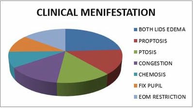
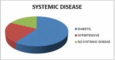
Introduction: Coronavirus had become an emergency in 2019. At that time invasive mucormycosis post-COVID-19 also emerged. There are multiple case reports and series describing mucormycosis in COVID-19. The recent emergence of the Coronavirus Disease (COVID-19) disease had been associated with reports of fungal infections such as aspergillosis and mucormycosis especially among critically ill patients like immune-compromised patients who have been treated with steroids. Method: A cross-sectional was conducted on patients with MRI and biopsy-proven mucormycosis with RT-PCR-confirmed COVID-19 from May to August 2021 at Dr D. Y. Patil Medical College and Research Institute, kadamwadi, Kolhapur, Maharashtra, India. the time interval between COVID-19 and mucormycosis, underlying systemic diseases, clinical features, course of the disease, and outcomes were collected and analyzed. (n=32). Result: out of 45 mucormycosis patients, 10 were female and 35 were male. The youngest participant was aged 12 years, and the oldest was 75 years old, the mean age was 53.33 ± 14.99 years. The majority of patients, 27 (60%) are diabetic and 10 are hypertensive (22.22%). 28 patients were COVID-19 positive. And 66.67% of patients were rhino orbital type of mucormycosis. All patients were treated with Amphotericin-B but out of 11(24.44%) got transcutaneous retrobulbar amphotericin –B injection. Conclusion: SARS-COV-2 infected patient numbers were increasing in COVOD -19 pandemic. Mucormycosis proved itself an epidemiological burden. So risk-based approach for patients with risk is necessary. Attention was to be given to that patients who were diabetic (uncontrolled sugar level), on combined use of steroids, or immune-modulating agents.

Keywords: Coronavirus, Rhino-Orbital mucormycosis, Aspergillosis

Corresponding Author How to Cite this Article To Browse
Milind Sabnis, Professor and HOD, Department of Ophthalmology, Dr D.Y. Patil Medical College Hospital and Research Institute, Kolhapur, Maharashtra, India.
Email:
Vaidehi Jethwa, Astha Vaghasia, Milind Sabnis, Increased Incidence of Rhino-Orbital Mucormycosis in an Educational Therapeutic Hospital During The Covid-19 Pandemic: An Observational Study. Trop J Ophthalmol Otolaryngol. 2023;8(2):6-10.
Available From
https://opthalmology.medresearch.in/index.php/jooo/article/view/246

Manuscript Received Review Round 1 Review Round 2 Review Round 3 Accepted
2023-06-07 2023-06-09 2023-06-16 2023-06-23 2023-06-30
Conflict of Interest Funding Ethical Approval Plagiarism X-checker Note
Nil Nil Yes 19%

© 2023by Vaidehi Jethwa, Astha Vaghasia, Milind Sabnisand Published by Siddharth Health Research and Social Welfare Society. This is an Open Access article licensed under a Creative Commons Attribution 4.0 International License https://creativecommons.org/licenses/by/4.0/ unported [CC BY 4.0].

Introduction

A Severe Acute Respiratory Syndrome Coronavirus 2 (SARS-CoV-2) infection is associated with a wide clinical spectrum of Coronavirus Disease 2019 (COVID-19) that ranges from being asymptomatic to a severe disease requiring intensive care unit (ICU) admission. [1] During 2nd wave of COVID-19 in India surge of mucormycosis cases was noted, especially in immune-compromised patients. Most commonly mucormycosis infection occurs due to inhalation of spores which affects body parts mostly pulmonary and cerebral infections. This fungus can survive well in glucose-rich media like and hence uncontrolled diabetic patients are more susceptible to this disease than patients with steroid treatment. [2] This fungus can be traced in the mucosa of the nasal and oral parts of a healthy human. The initial site of infection for this type of mucormycosis is the nasal turbinates, which further proliferate aggressively to involve the sinuses, palate, orbit, and brain. [3] this fungus is angioinvasive resulting in tissue ischemia and necrosis. [4] Along with the elastic tissues that form the outermost part of the blood vessels, a result of which the blood supply to the organs is obstructed. The infection spreads from the sinuses and gives rise to osteomyelitis bone, further, the infection spreads to the orbital structures and brain following the orbital route. [4] In contrast, in those who are immune‑compromised, the spores transform to hyphae; but since the white blood cells have lower efficacy on the hyphae, the fungi proliferate with greater ease. [5] Vision loss is due to the involvement of the optic nerve MRI was done to detect stage and spread of infection. Mucormycosis infection is typically confirmed by black tissue in the infected region while doing debridement. The manifestations of mucormycosis symptoms include:

facial pain, headache, inflammation, swelling of the periorbital and nasal region, bad odour, proptosis, eyelid drooping and edema, external and internal ophthalmoplegia, exophthalmos, nasal bleeding, facial paralysis accompanied by loss of vision, black nasal discharge. In case the disease progresses to the cranial region, the symptoms include lethargy, blindness, and seizures usually followed by death if the patient remains untreated.

Method

Type of study: A cross-sectional

Duration of study: from May to August 2021 at Dr D. Y. Patil Medical College and Research Institute, kadamwadi, Kolhapur, Maharashtra, India.

Sample size: A total of 45 patients with MRI and biopsy-proven mucormycosis with RT-PCR-confirmed COVID-19 during the above-given duration of the study.

Inclusion Criteria: Real-time reverse transcriptase polymerase chain reaction (RT-PCR) positive documented COVID-19 cases, histopathologic examination using hematoxyl and eosin (H&E) staining should be positive for Mucormycosis.

 OR

Computerized tomography (CT) scan of the orbit, paranasal sinuses, or Gadolinium-enhanced magnetic resonance imaging (MRI) of the orbit, paranasal sinuses, and brain should be confirmed as Mucormycosis.

Exclusion Criteria: Histopathologic examination not confirming Mucormycosis, computerised tomography (CT) scan of orbit, paranasal sinuses or Gadolinium-enhanced magnetic resonance imaging (MRI) of orbit, paranasal sinuses and brain not confirming Mucormycosis.

Data collection procedure: all previous history data like( ocular examination, age, sex, systemic disease, covid history, covid vaccine history, steroid usage history, loose tooth history, ventilator usage history) and all diagnosing data like ( MRI, CT- ORBIT, KOH mount, HistoPatho report, DNE (diagnostic nasal endoscopy ) collected by a junior resident of ophthalmology department and discussed with treating professor or Head of ophthalmology and ENT treatment data like ( amphotericin –B injection (IV and transcutaneous retrobulbar), FESS (functional endoscopic sinus surgery) surgery, Debridement surgery, tooth extraction surgery) achieved.

Methodology:

As part of ophthalmology checkups.

A total of 45 patients was taken for this study valid written consent was taken for the research and study, and detailed history was taken for past history, clinical history, drug history, and vaccination history, ocular examination was done on torch light followed by visual acuity will be checked, detailed anterior segment checked with a


slit lamp, ocular movements, sensation over the skin of sinuses, intraocular pressure checked, dilated fundoscopy done. 

opthal_246_01.JPG
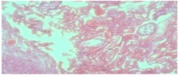
Figure 1: Histopathologically positive slide of mucormycosis given after debridement surgery.

opthal_246_02.JPG
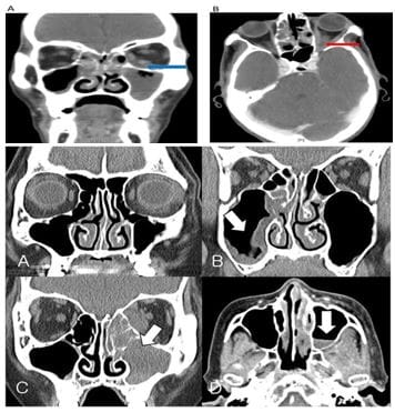
Figure 2: MRI showing mucormycosis in maxillary sinus and orbit.

Results

A total of 45 patents was taken in this study. According to systemic disease, they divided into 3 groups: 1) diabetic 60%, 2) hypertensive 22.22%, and 3) no systemic disease 17.77%.

Most patients like 28 (62.22%)are given the history of COVID -19 positive history and 5 patients (11.11%) took COVID vaccine of 2 dosages.

According to past history, a total of 17(37.77%) were given a history of steroid usage in the past. A total of 15 (33.33%) patients were on ventilators for more than 10 days. Total of 5 (11.11%) patients took ramdesiver (antiviral medication) in the past.

According to clinical manifestation 18(40%), patients came to OPD with both lid edema in eyes, 11 (24.44%) with proptosis, 10(22.22%) with ptosis, 10(22.22%) with congestion,7(15.55%) with chemosis and 8(17.77%) with fixed pupil and 11(24.44%) patients has extraocular movements restricted.

 According to treatment, all patients were given amphotericin – B Intravenous medication with 11 (24.44%) also given transcutaneous retrobulbar amphotericin – B injections and a total of 8 (17.77%) underwent tooth extraction surgery. All patients underwent debridement surgery to increase the healing process.

According to an outcome, a total of 41 patients were alive, 2 patients took discharged against medical advice and 2 patients were dead.

opthal_246_03.JPGopthal_246_04.JPG

Discussion

Mucormycosis is well-known as a black fungus which is highlighted during the COVID-19 second wave in India. Mucormycosis is a rare but potentially deadly fungal infection that affects individuals with weakened immune systems. The fungus responsible for the infection is known as Mucorales, and it is commonly found in soil, plants, and decaying organic matter. Mucormycosis typically develops in the sinuses, lungs, or brain and can cause severe damage if left untreated.


Mucormycosis is a rare but serious fungal infection that can cause great harm to individuals, especially to those with weakened immune systems. The fungal infection often affects the sinuses, brain, and lungs, leading to severe tissue damage and potentially life-threatening complications.

Iron theory, on the other hand, is a hypothesis that suggests that mucormycosis can be caused by iron overload in the body. Iron is an essential mineral that is important for many bodily functions, including oxygen transport, energy production, and immune system function. However, too much iron in the body can lead to an increase in free iron, which can promote the growth and spread of certain types of fungi, including mucormycetes. The iron theory suggests that reducing iron levels in the body could help prevent and treat mucormycosis. This can be done through various methods, including phlebotomy (bloodletting), chelation therapy (using chemicals to bind and remove excess iron), and dietary changes to limit iron intake. [8]

Overall, the iron theory remains a hypothesis, and more research is needed to determine its validity and potential efficacy for preventing and treating mucormycosis. However, it may provide a new direction for future studies on this serious fungal infection.

Classification of mucormycosis is according to the anatomic site of infection, reflecting in part the portals of entry in the human body. Spores enter the body either via the respiratory tract, through injured skin or via the percutaneous route (e.g. transmission of spores by contaminated needles or catheters), or ingestion of contaminated food. The disease may present in rhino-orbital-cerebral, pulmonary, cutaneous/subcutaneous, gastrointestinal or disseminated form. [7]

The symptoms of mucormycosis vary depending on the affected area of the body. If the infection is in the sinuses, symptoms may include nasal congestion, facial pain, and a black lesion on the nasal tissue. If the infection is in the lungs, symptoms may include chest pain, cough, fever, and shortness of breath. If the infection is in the brain, symptoms may include headache, fever, confusion, and seizures.

Mucormycosis is typically diagnosed through a combination of physical examination and laboratory tests, including CT scans, MRI

scans, and tissue biopsies. Early diagnosis and treatment are important to prevent the infection from spreading and causing more damage.

Treatment for mucormycosis typically involves antifungal medication, such as amphotericin B, which is administered intravenously. Other treatments may include surgery to remove infected tissue and improve ventilation, as well as supportive care to manage symptoms and reduce complications. Several risk factors make individuals more susceptible to developing mucormycosis. These include uncontrolled diabetes, leukaemia, organ or bone marrow transplantation, and prolonged use of corticosteroid medications.

Prevention of mucormycosis involves avoiding contact with mucorales, especially if an individual has a weakened immune system. This may include wearing protective clothing when gardening or doing outdoor activities, avoiding contaminated soil, and practising good hygiene to reduce the risk of infection.

Reference

01. Al-Tawfiq JA, Alhumaid S, Alshukairi AN, Temsah MH, Barry M, Al Mutair A, Rabaan AA, Al-Omari A, Tirupathi R, AlQahtani M, AlBahrani S. COVID-19 and mucormycosis superinfection: the perfect storm. Infection. 2021 Oct; 49:833-53. [Crossref][PubMed][Google Scholar]

02. Sundaram N, Bhende T, Yashwant R, Jadhav S, Jain A. Mucormycosis in COVID-19 patients. Indian Journal of Ophthalmology. 2021 Dec;69(12):3728. [Crossref][PubMed][Google Scholar]

03. Ajith Kumar AK, Gupta V. Rhino orbital cerebral mucormycosis. StatPearls. Treasure Island (FL): StatPearls Publishing; 2021. [Crossref][PubMed][Google Scholar]

04. Veisi A, Bagheri A, Eshaghi M, Rikhtehgar MH, Rezaei Kanavi M, Farjad R. Rhino orbital mucormycosis during steroid therapy in COVID 19 patients: A case report. Eur J Ophthalmol 2021; 11206721211009450. doi: 10. 1177/11206721211009450. Online ahead of print [Crossref][PubMed][Google Scholar]

05. Maini A, Tomar G, Khanna D, Kini Y, Mehta H, Bhagyasree V. Sino orbital mucormycosis in a COVID 19 patient: A case report. Int J Surg Case Rep 2021; 82:105957. . [Crossref][PubMed][Google Scholar]


06. Rudrabhatla PK, Reghukumar A, Thomas SV. Mucormycosis in COVID-19 patients: predisposing factors, prevention and management. Acta Neurologica Belgica. 2022 Apr 1:1-8. [Crossref][PubMed][Google Scholar]

07. Pillsbury HC, Fischer ND. Rhinocerebral mucormycosis. Archives of Otolaryngology. 1977 Oct 1;103(10):600-4. [Crossref][PubMed][Google Scholar]

08. Tabassum T, Araf Y, Moin AT, Rahaman TI, Hosen MJ. COVID-19-associated-mucormycosis: possible role of free iron uptake and immunosuppression. Molecular Biology Reports. 2022 Jan;49(1):747-54. [Crossref][PubMed][Google Scholar]