E-ISSN:2456-6454
P-ISSN:2581-4907
RNI:MPENG/2017/74152

Case Report

Pseudophakic bullous keratopathy

Tropical Journal of Ophthalmology and Otolaryngology

2022 Volume 7 Number 6 November-December
Publisherwww.medresearch.in

A late presented case of a pseudophakic bullous keratopathy with Descemet's membrane detachment treated by penetrating keratoplasty

Prasad N.1*, Dubey A.2, Kumar K.3
DOI: https://doi.org/10.17511/jooo.2022.i06.03

1* Nishi Prasad, Junior Resident, Third Year, Department of Ophthalmology, Gandhi Medical College, Bhopal, Madhya Pradesh, India.

2 Aditi Dubey, Associate Professor, Department of Ophthalmology, Gandhi Medical College, Bhopal, Madhya Pradesh, India.

3 Kavita Kumar, Professor & Head, Department of Ophthalmology, Gandhi Medical College, Bhopal, Madhya Pradesh, India.

Pseudophakic bullous keratopathy (PBK), also known as postoperative corneal edema, is the development of irreversible corneal edema after cataract surgery. Descemet’s membrane detachment (DMD) is also relatively common after cataract surgery. The purpose of this study is to present a case report on the visual outcome after penetrating keratoplasty in an elderly female with longstanding pseudophakic bullous keratopathy with descemet’s membrane detachment, who presented to cornea clinic with gradual progressive diminution of vision, five years after cataract surgery. Penetrating keratoplasty with a healthy donor cornea helped salvage her visual outcome.

Keywords: Pseudophakic bullous keratopathy, Descemet’s membrane detachment, Penetrating keratoplasty, Visual outcome

Corresponding Author How to Cite this Article To Browse
Nishi Prasad, Junior Resident, Third Year, Department of Ophthalmology, Gandhi Medical College, Bhopal, Madhya Pradesh, India.
Email:
Nishi Prasad, Aditi Dubey, Kavita Kumar, A late presented case of a pseudophakic bullous keratopathy with Descemet's membrane detachment treated by penetrating keratoplasty. Trop J Ophthalmol Otolaryngol. 2022;7(6):46-49.
Available From
https://opthalmology.medresearch.in/index.php/jooo/article/view/243

Manuscript Received Review Round 1 Review Round 2 Review Round 3 Accepted
2022-12-05 2022-12-07 2022-12-14 2022-12-21 2022-12-28
Conflict of Interest Funding Ethical Approval Plagiarism X-checker Note
Nil Nil Yes 17%

© 2022by Nishi Prasad, Aditi Dubey, Kavita Kumarand Published by Siddharth Health Research and Social Welfare Society. This is an Open Access article licensed under a Creative Commons Attribution 4.0 International License https://creativecommons.org/licenses/by/4.0/ unported [CC BY 4.0].

Introduction

Pseudophakic bullous keratopathy is characterized by epithelial and subepithelial bullae, stromal edema and endothelial decompensation through trauma secondary to cataract surgery especially phacoemulsification [1,4]. Initially, there is damage to the corneal endothelium which then progresses to stromal edema. This edema progress further to the subepithelial and epithelial layers of the cornea and results in bullae formation named bullous keratopathy [1,2]. Trauma to endothelium can occur during any intraocular surgery but bullous keratopathy is reported most commonly with phacoemulsification [1,3]. Descemet’s membrane detachment is most commonly caused by intraoperative trauma from instrument insertion into a corneal wound [5]. The most common cause of surgically induced DMD is cataract surgery [6]. As DMD prolongs, the cornea becomes edematous due to endothelial dysfunction [6]. Most of the cases of DMD are small peripheral detachments at the site of corneal incision and are clinically insignificant but some of the cases are large, involving the central cornea and those requiring a corneal transplant to regain corneal clarity [5]. As advancements in surgical techniques have evolved with newer IOL designs and better training of ophthalmologists, the incidence has reduced drastically [1]. However, it remains an important cause of visual morbidity after a routine and complicated cataract surgery [1].

Case Report

A sixty-six years old female presented with a diminution of vision in the left eye for five years, which was gradual in onset and progressive. She had been operated on for both eye cataract surgery five years back. She had no history of any chronic illness. The best corrected visual acuity in the right eye was 6/6 and in the left eye was 1/60. The right eye anterior segment was normal. Left eye cornea revealed multiple bullae in the epithelium, diffuse stromal edema, descemet’s membrane detachment, the pupil was mid-dilated non reacting to light, PCIOL in situ (fig 1). Intraocular pressure was 18 and 20mmHg in right and left eye respectively. Fundus under mydriasis showed a faintly visible disc and blood vessels in the left eye and within the physiological limit in the right eye.

Anterior segment OCT of the left eye confirmed descemet’s membrane detachment (fig 2). The diagnosis of pseudophakic bullous keratopathy with descemet’s membrane detachment was established. Since it was a very late presentation, penetrating keratoplasty was done in the left eye from a suitable healthy donor. The host cornea was removed and the donor cornea was opposed to the host with the help of 16 interrupted 10-0 nylon sutures.

Following a penetrating keratoplasty, oral and topical antibiotic, topical steroids, cycloplegic, hyperosmotic eye drops and lubricants were given. Oral acetazolamide was also given in the first week and intraocular pressure was monitored properly. Good postoperative care is extremely important in these cases for graft survival and visual rehabilitation. Her visual outcome started improving. Graft clarity was 1+ with healthy graft host junction (fig 3 and fig 4). The best corrected visual acuity was improved to 6/9 in two weeks.

opthal_243_01.JPG
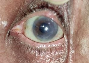
Fig 1: left eye cornea showing pseudophakic bullous keratopathy

opthal_243_02.JPG
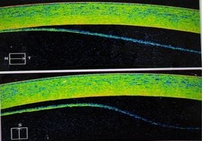
Fig 2: Left eye anterior segment OCT showing Descemet's membrane detachment


opthal_243_03.JPG
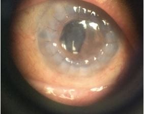
Fig 3: Post-penetrating keratoplasty after 1 week with healthy graft

opthal_243_04.JPG
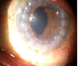
Fig 4: Post-penetrating keratoplasty after 2 weeks with healthy graft

Discussion

Pseudophakic bullous keratopathy is due to endothelial loss after surgical trauma, especially in the elderly [1,4]. The other causes are endothelial burns due to thermal damage secondary to high energy use during phacoemulsification. High irrigation and aspiration can also damage endothelium during cataract surgery [1,4]. PBK can also occur after excessive use of ultrasound energy during phacoemulsification, complicated cataract surgery, anterior vitrectomy close to the endothelium, and during nucleus delivery in manual small incision cataract surgery due to nucleus rub over cornea [1,4]. The epithelium and endothelium act as a barrier to water and electrolytes owning to their semi-permeable nature of the membrane.

Na+-K+-ATPase pump in the endothelium prevents corneal hydration and helps in maintaining transparency. Any damage to endothelial cells hampers corneal transparency. Surgically induced trauma, excessive ultrasound energy use in surgery, persistent inflammation and endothelial dystrophies can promote cell loss [1]. When the cell density reaches a critically low value, it results in the development of pseudophakic bullous keratopathy. The pump fails and manifests as stromal edema, which can change in response to intraocular pressure. The aqueous migrates from stroma to epithelium resulting in epithelial edema, blisters and bullae formation. Descemet’s membrane detachment is a serious complication after surgical procedures involving anterior chamber manipulation [7]. Rarely it can occur in the intermediate or late postoperative period after uncomplicated surgery [7]. Of all the procedures involving anterior chamber entry, DMD is reported most commonly after cataract surgery [7]. There are two types of DMD: peripheral and central. Peripheral DMD is small with minimal corneal edema and therefore given conservative management. Central DMD or involving the visual axis requires surgical intervention. Delayed onset DMD should be considered as one of the differentials in cases with late-onset corneal edema post-cataract surgery [7]. Our patient presented late with a gradually progressive diminution of vision after cataract surgery. Penetrating keratoplasty is the gold standard surgery in pseudophakic bullous keratopathy [1].

In this surgery, a full thick host cornea is replaced by a donor cornea which is opposed to the host cornea with interrupted or continuous sutures. Penetrating keratoplasty can salvage vision in late-presented or neglected cases of PBK with DMD. Patient education also plays an important role in managing the case. The patient should be educated regarding the pathology and mechanism behind corneal edema and also regarding the management options.

Conclusion

Pseudophakic bullous keratopathy is one of the common ocular pathology in clinical practice after cataract surgery. However, the incidence has reduced but it remains one of the common causes of corneal transplantation worldwide.


Descemet’s membrane detachment is also relatively common after cataract surgery. Prompt diagnosis and management with expert hands can safeguard visual outcomes in the majority of cases. In late presentation or neglected cases, penetrating keratoplasty can help in salvaging vision.

Reference

01. Gurmani B, Kaur K. Pseudophakic Bullous Keratopathy. [Updated 2022 Dec 6]. In: StatsPearls. Treasure Island (FL): StatPearls Publishing; 2022 Jan [Crossref][PubMed][Google Scholar]

02. Claesson M, Armitage WJ, Stenevi U. Corneal oedema after cataract surgery: predisposing factors and corneal graft outcome. Acta Ophthalmol. 2009 Mar;87(2):154-9. [Crossref][PubMed][Google Scholar]

03. Feizi S. Corneal endothelial cell dysfunction: etiologies and management. Ther Adv Ophthalmol. 2018 Jan-Dec;10:2515841418815802. [Crossref][PubMed][Google Scholar]

04. Pricopie S, Istrate S, Voinea L, Leasu C, Paun V, Radu C. Pseudophakic bullous keratopathy. Rom J Ophthalmol. 2017 Apr-Jun;61(2):90-94. [Crossref][PubMed][Google Scholar]

05. Samarawickrama C, Beltz J, Chan E. Descemet’s membrane detachments post cataract surgery: a management paradigm. Int J Ophthalmol. 2016;9(12):1839-1842. Published 2016 Dec 18. doi:10.18240/ijo.2016.12.23 [Crossref][PubMed][Google Scholar]

06. Wang SW, Tseng SH. Occult Descemet’s membrane detachment after phacoemulsification surgery mimicking pseudophakic bullous keratopathy. Taiwan J Ophthalmol. 2015;5(3):136-139. doi:10.1016/j.tjo.2015.04.001 [Crossref][PubMed][Google Scholar]

07. Bhatia, H. K. , Gupta, R. Delayed-onset descemet’s membrane detachment after uneventful cataract surgery treated by corneal venting incision with air tamponade: a case report. BMC Ophthalmol 16, 35 (2016). doi:10.1186/s12886-016-0212-6 [Crossref][PubMed][Google Scholar]