E-ISSN:2456-6454
P-ISSN:2581-4907
RNI:MPENG/2017/74152

Research Article

Atropine

Tropical Journal of Ophthalmology and Otolaryngology

2021 Volume 6 Number 6 November December
Publisherwww.medresearch.in

Can Myopia Be Reversed? - A Study on the Role of Atropine Eye drops in Arresting Myopia Progression

Tripathi A.1*, Gupta R.2, Mohan S.3
DOI: https://doi.org/10.17511/jooo.2021.i06.02

1* Anchal Tripathi, Senior Resident, Department of Ophthalmology, GSVM Medical College, Kanpur, U.P, India.

2 RC Gupta, Professor and Head, Department of Ophthalmology, LLRM, Meerut, U.P, India.

3 Shalini Mohan, Associate Professor, Department of Ophthalmology, GSVM Medical college, Kanpur, U.P, India.

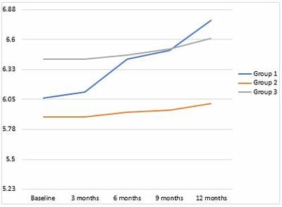
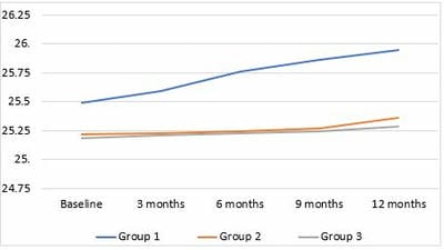
Introduction: Atropine is being explored as a potent agent for arresting myopia progression for ages. In our study, we attempt to investigate the efficacy of two different concentrations of atropine in halting myopia progression in Indian eyes. Objectives: 1. To investigate the effect of atropine in retarding progression of myopia. 2. To compare rates of retardation of myopia progression using 0.05% atropine and 0.1% atropine. Methods: Patients were selected from children between 6-18 yrs of age, visiting the outpatient department of our hospital, with spherical equivalent (SE) =>-1D in both the eyes and spherical equivalent (SE) progression rate >= 0.5D/year. Groups with 0.05% atropine eye drop, 0.1% atropine eye drops and control were allocated randomly. Myopia progression was measured by the change in spherical equivalent and axial length at baseline and every three months till one year. Results: In our study, a total of 48 eyes of 24 children were included, out of which 83.33% were in the age group 6-12years. The mean change in spherical equivalent in our study after one year was -0.72±0.21D, -0.11±0.096D and -0.19±0.18D in control, 0.1% and 0.05% atropine groups, respectively. Also, the mean change in axial length of our study after one year was 0.45±0.15mm, 0.13±0.20mm and 0.11±0.02mm in control, 0.1% and 0.05% atropine groups, respectively. Changes in both parameters were found to be statistically significant. Conclusion: We conclude that night-time application of 0.05% and 0.1% atropine eye drops is efficacious in retarding progressive myopia in Indian eyes.

Keywords: Atropine, Axial length, Myopia, Spherical equivalent

Corresponding Author How to Cite this Article To Browse
Anchal Tripathi, Senior Resident, Department of Ophthalmology, GSVM Medical College, Kanpur, U.P, India.
Email:
Anchal Tripathi, RC Gupta, Shalini Mohan, Can Myopia Be Reversed? - A Study on the Role of Atropine Eye drops in Arresting Myopia Progression. Trop J Ophthalmol Otolaryngol. 2021;6(6):114-120.
Available From
https://opthalmology.medresearch.in/index.php/jooo/article/view/217

Manuscript Received Review Round 1 Review Round 2 Review Round 3 Accepted
2021-10-16 2021-10-18 2021-10-25 2021-11-01 2021-11-08
Conflict of Interest Funding Ethical Approval Plagiarism X-checker Note
Nil Nil Yes 17%

© 2021by Anchal Tripathi, RC Gupta, Shalini Mohanand Published by Siddharth Health Research and Social Welfare Society. This is an Open Access article licensed under a Creative Commons Attribution 4.0 International License https://creativecommons.org/licenses/by/4.0/ unported [CC BY 4.0].

Introduction

The prevalence of myopia has increased all over the world drastically over the past decade [1]. Myopia prevalence in young children of South Asia has risen to 95.5% [2,3]. Studies across Europe have also shown a rising tendency of myopia among schooling children [4]. These increasing trends also include the increasing prevalence of high myopia (<-6D; axial length>25mm). High myopia may lead to myopic macular degeneration, foveal schisis and glaucoma.2 There is a 30% absolute risk of severe visual impairment in individuals with an axial length of 30mm or more [5].

Currently, treatment options for progressive myopia are conservative and pharmacological interventions [6]. The results of the conservative regimens, except for orthokeratology, are comparatively unsatisfactory [7]. Pharmacological intervention has been higher efficacy, especially atropine eye drops [8].

Atropine is a non-selective muscarinic receptor antagonist (M-antagonist). It has been widely used in ophthalmology for ages as a cycloplegia inducing pupil dilator. In 1864, Donders was the first to suggest the usage of atropine as a pharmacological treatment for progressive myopia [9]. In 2012, Chia et al. concluded that daily application of 0.1% and 0.25% atropine was well-tolerated, and therefore, recommended for children with progressive myopia [10]. However, there are limited studies on the potency of atropine on Indian eyes.

The mechanism by which atropine retards the progression of myopia is still unknown. Some authors suggest that the action of atropine on the retinal and the scleral receptors leads to reshaping the sclera, thus arresting the growth of the eyeball [11]. There is another theory that proposes that the pupil dilation caused by atropine leads to increased ultraviolet exposure, which consequently may cause the collagen bonds in the sclera to break and cross-link, and therefore, curbing scleral growth [12].

Material And Methods

Setting Of Study- Hospital based.

Type Of Study- a prospective study.

Duration Of Study- 2 years

Sampling Methods-

Patients were selected randomly from patients visiting the outpatient department of Ophthalmology, LLRH, GSVM Medical College, Kanpur

Sample Size Calculation- The sample size is calculated using the following formula:

n = (Z1-α/2− Z1-β/2)2 SD2/d2

Z1-α/2= Level of significance

Z1-β/2= Power of the study

SD= Standard deviation

d = Effect size

Inclusion Criteria

  1. Patients whose parents or legal guardians consent to participate in the study.
  2. Patients between 6-18 years of age.
  3. Patients with spherical equivalent (SE) =>-1D.
  4. Patients with SE progression rate >= 0.5D/year.

Exclusion Criteria

  1. Uncooperative patients/legal guardians.
  2. Patients with myopia related to retinal dystrophies or collagen syndromes or any posterior segment disease.
  3. Patients with any anterior segment disease.
  4. Patients with amblyopia or any ocular disease other than refractive error.
  5. Patients less than six years of age.
  6. Patients with myopia < -1D.
  7. Patients with SE<0.5D/yr.

Data Collection Procedure: Three groups were allocated on a 1:1:1 basis.

Group 1: controls given placebo (carboxymethylcellulose 5%) eye drops.

Group 2: children who were given 0.1% atropine eye drops.

Group 3: children who were given 0.05% atropine eye drops.

Myopia progression was measured by the change in spherical equivalent (SE) and axial length (AL) at baseline and every three months till one year.

SE was calculated using the standard formula: (SE=sphere+1/2cylinder)


Ethical Consideration and Permission: The study was approved by the ethical review committee (Institutional Review Board) of our institution. It was conducted following Good Clinical Practice within Helsinki’s Declaration of 1975, as revised in 2000. The patients/subjects were selected after getting an informed consent signed by their parents.

Statistical Analysis

  • Statistical analysis was performed using SPSS 21.0 (IBM SPSS Inc., Chicago, IL, USA) for Windows statistical package.
  • The unpaired t-test was used as a test of significance to compare variables between two groups. The paired t-test was used to compare pre-and post-treatment progression.
  • Probabilities were two-tailed and considered statistically significant if p<0.05.

Results

In our study, 48 eyes of 24 children were included, out of which 20 (83.33%) were in the age group 6-12years, whereas 4 (16.67%) were in the 13-18years age group. Also, 13 (54.17%) children were males compared to 11 (45.83%) females.

The mean change in spherical equivalent in our study after one year was -0.72±0.21D, -0.11±0.096D and -0.19±0.18D in placebo, 0.1% and 0.05% atropine groups, respectively (FIGURE 1). Also, the mean change in axial length of our study after one year was 0.45±0.15mm, 0.13±0.20mm and 0.11±0.02mm in placebo, 0.1% and 0.05% atropine groups, respectively (FIGURE 2). Changes in both parameters were found to be statistically significant. However, differences in myopia progression (0.19 D) and axial length change (0.14 mm) between groups were small and clinically insignificant.

Out of the total 39 children included, the total number of children excluded from the study was 15(51.72%).

Five patients from group 2 (0.1% atropine eye drops) and one patient from group 3 (0.05% atropine eye drops) were excluded from the study due to complaints of blurring of near vision and hence, difficulty in studying. Three patients from group 2 (0.1% atropine eye drops) and two patients from group 3 (0.05% atropine

eye drops) were excluded from the study due to unendurable photosensitivity. Four patients from group 2 developed mild allergic reactions to the eyedrops and, hence, were excluded from the study. Therefore, the most common side-effect encountered in this study was blurring of near vision (Figure 3A).

Out of 15 children, who experienced adverse effects due to atropine eye drops, 12(80%) children were in the 13-18 years age group, while only 3(20%) were in the 6-12 years age group  (Figure 3B).

opthal_217_02.JPG
Figure 1: Progression Of Spherical Equivalent In 1 Year

opthal_217_03.JPG
Figure 2: Progression Of Axial Length In 1 Year (In Mm)


Figure 3 a: adverse effects (in both grpoups)


Figure 3b : adverse effects in different age groups

Discussion

Walline et al. [6] did a Cochrane database systematic review on interventions to slow myopia progression in children and concluded that the anti-muscarinic topical medications are most effective in controlling myopia progression. A similar evidence-based review was carried out by Saw et al. [11]. With similar conclusions. Therefore, the effectiveness of different anti-muscarinic agents is being studied worldwide to cease the progression of this refractive error [8,10,13,15,19].

In our study, the population included was entirely Indian. Kothari et al. [13] also studied the efficacy of atropine in myopia control in the Indian people, but the concentration used was 1%. It has been suggested that atropine may have different potency in the scenario of Indian eyes since the density and the distribution of these receptors differ significantly in differently pigmented eyes [13,14].

The mean change in spherical equivalent in our study after one year was -0.72±0.21D, -0.11±0.096D and -0.19±0.18D in placebo, 0.1% and 0.05% atropine groups, respectively, which concurred with the findings in the Low-concentration for myopia progression (LAMP) study by JC Yam et al. [15]. They found out that the mean change in the spherical equivalent of refraction was -0.27±0.61D, -0.46±0.45D, -0.59±0.61D and -0.81±0.53D in the 0.05%, 0.025% and 0.01% atropine groups and placebo groups, respectively [15].

The mean change in axial length of our study after one year was 0.45±0.15mm, 0.13±0.20mm and 0.11±0.02mm in placebo, 0.1% and 0.05% atropine groups, respectively.

JC Yam et al. [14] also found similar results in axial length progression. The mean increase in axial length was 0.20±0.25mm, 0.29±0.20mm, 0.36±0.29mm and 0.41±0.22mm in the 0.05%, 0.025% and 0.01% atropine groups and placebo groups, respectively.

In the present study, change in spherical equivalent, and axial length was found to be significant both for 0.1% atropine eye drops as well as 0.05% atropine eye drops as compared to placebo eye drops (p<0.01). However, these differences between groups were statistically insignificant (p>0.05). This is similar to results obtained by Chia et al. [10], who did a five year clinical trial on Atropine to treat myopia (ATOM) 2. The mean myopia progression at 2 years was −0.30±0.60, −0.38±0.60, and −0.49±0.63 D in the atropine 0.5%, 0.1%, and 0.01% groups, respectively (P=0.02 between the 0.01% and 0.5% groups; between other concentrations P > 0.05). The mean increase in axial length was 0.27±0.25, 0.28±0.28, and 0.41±0.32 mm in the 0.5%, 0.1%, and 0.01% groups, respectively (P < 0.01 between the 0.01% and 0.1% groups and between the 0.01% and 0.5% groups). However, differences in myopia progression (0.19 D) and axial length change (0.14 mm) between groups were small and clinically insignificant.

Only those patients who did not miss or stop the atropine eye drops during the study period were included for the statistical analysis. Out of the total 29 children had, the total number of children excluded from the study was 15 (51.72%).

Five patients (excluded from the study) stopped using 0.1% atropine eye drops, and one patient (excluded from the study) stopped using 0.05% atropine eye drops due to blurring of near vision, leading to trouble in doing near work. Three patients (excluded from the study) stopped using 0.1% atropine eye drops, and two patients (excluded from the study) stopped using 0.05% atropine eye drops because of unendurable photosensitivity despite prescription of photochromatic glasses, four patients (excluded from the study) developed an allergy to 0.1% atropine. Therefore, the most common side-effect encountered in our research was the blurring of near vision. This is in contrast to results obtained by Chia et al. [10], who observed allergic conjunctivitis and dermatitis as the most common side effects,


with 16 cases in the 0.1% and 0.5% atropine groups and no cases in the 0.01% group. Although there was no history of fever or rashes over the face in our study, the instructions to occlude puncta after instilling the eye drops were given to reduce the risk of systemic side effects.

Out of 15 children, who experienced adverse effects due to atropine eye drops, 80% of children were in the 13-18 years age group, which suggests that the use of atropine eye drops to arrest the progression of myopia in the adolescent age group might be more risky than beneficial. To our knowledge, there is no other study that compares the adverse effects of atropine eye drops in different age groups.

Tong et al. [16] reported rebound myopia progression after stopping treatment. Similar results were obtained by Huang et al. [17] and Chia et al. [18]. Nonetheless, absolute myopia progression after three years was found to be considerably less in the atropine group than in the control group.

The mechanism by which atropine retards the progression of myopia is still unknown. Some authors suggest that the action of atropine on the retinal and the scleral receptors leads to reshaping the sclera, thus arresting the growth of the eyeball [11,20,21]. There is another theory that proposes that the pupil dilation caused by atropine leads to increased ultraviolet exposure, which consequently may cause the collagen bonds in the sclera to break and cross-link, and therefore, curbing scleral growth [12].

Limitations

  • The study population is very small.
  • Long term follow-up of the patients might be required to confirm the long term outcome of the study.

Conclusion

Once-daily bedtime application of 0.05% and 0.1% atropine eye drops was efficacious in retarding progressive myopia in Indian eyes.

Dropout rate was more in the adolescent population, which may be due to more near work. The risk versus benefit ratio of usage of atropine eye drops in this population was higher due to more adverse effects.

More studies are required to determine the earliest age for atropine eye drops for myopia.

Recommendations: By our study, we recommend that 0.05% atropine should be used as early as possible, preferably upon the diagnosis of myopia, in young children. Once-daily nighttime application is recommended.

What Our Study Adds To the Existing Knowledge

  1. Atropine eye drops should be initiated early, as soon as myopia is detected in a child.
  2. Higher concentrations of atropine eyedrops are recommended in the pre-adolescent age group, that is, 0.1% as compared to 0.01% that is being used worldwide.
  3. Once the child enters adolescence, usage of atropine for arresting myopia is almost ineffective.

Contribution of Authors: Dr (Capt) Anchal Tripathi: Data collection, analysis and manuscript preparation, Dr RC Gupta: Concept and study design, Dr Shalini Mohan: Concept and statistical analysis.

Reference

01. Morgan IG, Ohno-Matsui K, Saw SM. Myopia. Lancet. 2012 May 5;379(9827):1739-48. doi: 10.1016/S0140-6736(12)60272-4 [Crossref][PubMed][Google Scholar]

02. Pan CW, Ramamurthy D, Saw SM. Worldwide prevalence and risk factors for myopia. Ophthalmic Physiol Opt. 2012 Jan;32(1):3-16. doi: 10.1111/j.1475-1313.2011.00884.x [Crossref][PubMed][Google Scholar]

03. Sun J, Zhou J, Zhao P, Lian J, Zhu H, Zhou Y, et al. High prevalence of myopia and high myopia in 5060 Chinese university students in Shanghai. Invest Ophthalmol Vis Sci. 2012 Nov 1;53(12):7504-9. doi: 10.1167/iovs.11-8343 [Crossref][PubMed][Google Scholar]

04. Williams KM, Bertelsen G, Cumberland P, Wolfram C, Verhoeven VJ, Anastasopoulos E, et al. Increasing Prevalence of Myopia in Europe and the Impact of Education. Ophthalmology. 2015 Jul;122(7):1489-97. doi: 10.1016/j.ophtha.2015.03.018 [Crossref][PubMed][Google Scholar]


05. Vongphanit J, Mitchell P, Wang JJ. Prevalence and progression of myopic retinopathy in an older population. Ophthalmology. 2002 Apr;109(4):704-11. doi: 10.1016/s0161-6420(01)01024-7 [Crossref][PubMed][Google Scholar]

06. Walline JJ, Lindsley K, Vedula SS, Cotter SA, Mutti DO, Twelker JD. Interventions to slow progression of my-opia in children. Cochrane Database Syst Rev. 2011 Dec 7;(12):CD004916. doi: 10.1002/14651858.CD004916 [Crossref][PubMed][Google Scholar]

07. Sun Y, Xu F, Zhang T, Liu M, Wang D, Chen Y, et al. Orthokeratology to control myopia progression: a me-ta-analysis. PLoS One. 2015 Apr 9; 10(4):e0124535. doi: 10.1371/journal.pone.0124535 [Crossref][PubMed][Google Scholar]

08. Gwiazda J. Treatment options for myopia. Optom Vis Sci. 2009 Jun;86(6):624-8. doi: 10.1097/OPX.0b013e3181a6a225 [Crossref][PubMed][Google Scholar]

09. Donders, Franciscus Cornelis, and William Daniel Moore. On the anomalies of accommodation and refrac-tion of the eye: With a preliminary essay on physiological dioptrics. Vol. 22. New Sydenham Society, 1864 [Crossref][PubMed][Google Scholar]

10. Chia A, Chua WH, Cheung YB, Wong WL, Lingham A, Fong A, et al. Atropine for the treatment of child-hood myopia: safety and efficacy of 0. 5%, 0. 1%, and 0. 01% doses (Atropine for the Treatment of Myopia 2). Ophthalmology. 2012 Feb;119(2):347-54. doi: 10.1016/j.ophtha.2011.07.031 [Crossref][PubMed][Google Scholar]

11. Saw SM, Shih-Yen EC, Koh A, Tan D. Interventions to retard myopia progression in children: an evidence-based update. Ophthalmology. 2002 Mar;109(3):415-21; discussion 422-4; quiz 425-6, 443. doi: 10.1016/s0161-6420(01)00972-1 [Crossref][PubMed][Google Scholar]

12. Prepas SB. Light, literacy and the absence of ultraviolet radiation in the development of myopia. Med Hy-potheses. 2008;70(3):635-7. doi: 10.1016/j.mehy.2007.07.023 [Crossref][PubMed][Google Scholar]

13. Kothari M, Rathod V. Efficacy of 1% atropine

eye drops in retarding progressive axial myopia in Indian eyes. Indian J Ophthalmol. 2017 Nov;65(11):1178-1181. doi: 10.4103/ijo.IJO_418_17 [Crossref][PubMed][Google Scholar]

14. Salazar M, Shimada K, Patil PN. Iris pigmentation and atropine mydriasis. J Pharmacol Exp Ther. 1976 Apr;197(1):79-88. [Crossref][PubMed][Google Scholar]

15. Anderson HA, Bertrand KC, Manny RE, Hu YS, Fern KD. Comparison of two drug combinations for dilat-ing dark irides. Optom Vis Sci. 2010 Feb;87(2):120-4. doi: 10.1097/OPX.0b013e3181cc8da3 [Crossref][PubMed][Google Scholar]

16. Tong L, Huang XL, Koh AL, Zhang X, Tan DT, Chua WH. Atropine for the treatment of childhood myo-pia: effect on myopia progression after cessation of atropine. Ophthalmology. 2009 Mar;116(3):572-9. doi: 10.1016/j.ophtha.2008.10.020 [Crossref][PubMed][Google Scholar]

17. Huang J, Wen D, Wang Q, McAlinden C, Flitcroft I, Chen H, et al. Efficacy Comparison of 16 Interventions for Myopia Control in Children: A Network Meta-analysis. Ophthalmology. 2016 Apr;123(4):697-708. doi: 10.1016/j.ophtha.2015.11.010 [Crossref][PubMed][Google Scholar]

18. Chia A, Chua WH, Wen L, Fong A, Goon YY, Tan D. Atropine for the treatment of childhood myopia: changes after stopping atropine 0. 01%, 0. 1% and 0. 5%. Am J Ophthalmol. 2014 Feb;157(2):451-457.e1. doi: 10.1016/j.ajo.2013.09.020 [Crossref][PubMed][Google Scholar]

19. Siatkowski RM, Cotter S, Miller JM, Scher CA, Crockett RS, Novack GD; US Pirenzepine Study Group. Safety and efficacy of 2% pirenzepine ophthalmic gel in children with myopia: a 1-year, multicenter, double-masked, placebo-controlled parallel study. Arch Ophthalmol. 2004 Nov;122(11):1667-74. doi: 10.1001/archopht.122.11.1667 [Crossref][PubMed][Google Scholar]

20. Tigges M, Iuvone PM, Fernandes A, Sugrue MF, Mallorga PJ, Laties AM, Stone RA. Effects of muscarinic cholinergic receptor antagonists on postnatal eye growth of rhesus monkeys. Optom


Vis Sci. 1999 Jun;76(6):397-407. doi: 10.1097/00006324-199906000-00020 [Crossref][PubMed][Google Scholar]

21. Lind GJ, Chew SJ, Marzani D, Wallman J. Muscarinic acetylcholine receptor antagonists inhibit chick scleral chondrocytes. Invest Ophthalmol Vis Sci. 1998 Nov;39(12):2217-31. [Crossref][PubMed][Google Scholar]