E-ISSN:2456-6454
P-ISSN:2581-4907
RNI:MPENG/2017/74152

Research Article

Donor

Tropical Journal of Ophthalmology and Otolaryngology

2021 Volume 6 Number 4 July-August
Publisherwww.medresearch.in

Effect of donor and host factors on corneal graft transparency

Jain A.1*, Joshi H.2, Jain N.3
DOI: https://doi.org/10.17511/jooo.2021.i04.01

1* Amisha Jain, Assistant Professor, Department of ophthalmology, S.B.KS. M.I and R.C., Dhiraj General Hospital, Piparia, Gujrat, India.

2 Hema Joshi, Cataract, Cornea and Ocular surface, H V Desai Eye Hospital, Pune, Maharashtra, India.

3 Nimish Jain, Assistant Professor, Department of Cardiac Anaesthesia, S.B.KS. M.I and R.C., Dhiraj General Hospital, Piparia, Gujrat, India.

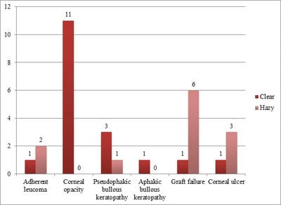
Objective: To study the correlation between donor factors and recipients' factors on graft clarity. Materials and methods: The study comprised 30 cases of Keratoplasty surgery with a follow up of 6 months. All donor corneas were evaluated by Konan specular microscope for endothelial cell count; details of the donor like age, cause of death were noted. The patients were divided into two groups, Group 1 had graft failure, and Group 2 had clear corneas. Observation and Result: There were 12 patients in group 1 and 18 patients in group 2 with six months of follow up. The mean endothelial cell count in group 1 was 1942.3/mm2, and group 2 was 2334.8/mm2. There is a significant difference in the mean endothelial cell count between the two groups. On analysing the indication for Keratoplasty in two groups, the outcome was best for the corneal opacity group during worst for the graft failure group. Conclusion: Donor endothelial cell count significantly influenced graft outcome; rest donor factors (age, death enucleation interval, enucleation surgery interval) don't affect graft survival. Indication for Keratoplasty is a significant predictor of graft survival.

Keywords: Donor factors, Endothelial cell count, Keratoplasty, Graft failure, Indication for Keratoplasty, Host factors

Corresponding Author How to Cite this Article To Browse
Amisha Jain, Assistant Professor, Department of ophthalmology, S.B.KS. M.I and R.C., Dhiraj General Hospital, Piparia, Madhya Pradesh, India.
Email:
Jain A, Joshi H, Jain N. Effect of donor and host factors on corneal graft transparency. Trop J Ophthalmol Otolaryngol. 2021;6(4):56-61.
Available From
https://opthalmology.medresearch.in/index.php/jooo/article/view/206

Manuscript Received Review Round 1 Review Round 2 Review Round 3 Accepted
2021-07-03 2021-07-13 2021-07-23 2021-07-30 2021-08-09
Conflict of Interest Funding Ethical Approval Plagiarism X-checker Note
No Nil Yes 9%

© 2021 by Amisha Jain, Hema Joshi, Nimish Jain and Published by Siddharth Health Research and Social Welfare Society. This is an Open Access article licensed under a Creative Commons Attribution 4.0 International License https://creativecommons.org/licenses/by/4.0/ unported [CC BY 4.0].

Introduction

Blindness is a significant public health problem in developing countries. Cataracts and corneal diseases are the two major causes of blindness in countries in developing economies. [1]. According to the World Health Organization, corneal diseases are one of the significant causes of vision loss and blindness in the world today, after cataracts and glaucoma. [2].

In India, it is estimated that there are approximately 6.8 million people with vision less than 6/60 in at least one eye due to corneal diseases; out of these, about a million have bilateral involvement. [3,4]. It is estimated that the number of people with unilateral corneal blindness in India will increase to 10.6 million by 2020. [4].

According to the National Programme for Control of Blindness (NPCB) estimates, about 120,000 corneal blind persons in the country. According to a recent estimate, there is an addition of 25,000-30,000 corneal blindness cases every year in the country. [5]. The burden of corneal disease in India is reflected by 90% of the global cases of ocular trauma, and corneal ulceration leading to corneal blindness occur in developing countries. [6].

The importance of corneal transplant lies in the fact that successful transplantation can restore excellent sight to corneal blindness. Penetrating Keratoplasty is the most frequently performed and the most successful form of transplantation. [7]. The proportion of corneal blind patients who could derive long term benefits from corneal grafting depends on the graft transparency. Trans­parency is affected by many factors.

These factors are the type of donor material, condition of the recipient's eye, the operative procedures and postoperative treatment. The quality of donor material depends upon the donor's age, cause of death, the interval between the time of death and time of enucleation, the duration for which the donor's eye was preserved, the clinical condition of the donor's vision, its bacteriological and method of preservation and endothelial cell count.

The function of Endothelial cells of the cornea maintains this tissue in a dehydrated state by their pumping activity, assuring its transparency. The corneal endothelium consists of a single layer of non-regenerating, mainly hexagonal cells.

It plays a pivotal role in maintaining a clear cornea by securing a state of relative dehydration of the corneal stroma The factors related to the recipient's eye are the cause and extent of corneal pathology, corneal vascularisation and other associated ocular pathology. The operative factors which can affect the graft transparency are the size of the graft, the suturing technique used, the operative technique and the complications. Postoperative complications and postoperative treatment are essential factors that govern the transparency of graft.

Graft Failure: Graft failure refers to any graft that does not retain its transparency sufficient for adequate vision. There are multiple causes of graft failure, some of which are mentioned, and others may be immunological. Primary graft failure: It is defined as corneal oedema that is present right from the time of Keratoplasty, does not clear, and is not associated with rejection or secondary cause of graft failure. It may manifest on the first postoperative day. Corneal transplantation is the most frequent and successful transplant. The remarkable survival of corneal transplants can be primarily attributed to their unique avascular structure; it makes the cornea is immune privileged. Immune-mediated rejection is the leading cause of graft failure; up to 30% of penetrating Keratoplasty will have at least one episode of rejection.

Materials and methods

Study design: Prospective interventional study.

The study was conducted in the department of ophthalmology tertiary care centre of Central India. The present study comprised 30 cases of Keratoplasty surgery with a follow up of 6 months. The patients were divided into two groups; Group 1 had graft failure, Group 2 had clear corneas. All donor corneas were evaluated by Konan specular microscope for endothelial cell count; details of the donor like age, cause of death were noted. Death enucleation interval and death transplantation interval was noted. All corneas were preserved in M.K. media, and patients undergoing Keratoplasty were evaluated.

Inclusion criteria: Only those corneas which were procured within 6 hours of death are used.

Patients with at least six months of follow up post keratoplasty.

Exclusion criteria: Patients who lost follow up.


Age less than ten years.
Corneal Graft Rejection assessed by

Symptoms

  • Diminution of vision, pain, redness and photophobia after keratoplasty surgery

Signs

  • The white line on the corneal endothelium
  • Stromal thickening edema or infiltrates
  • Subepithelial or epithelial edema
  • Conjunctival injection
  • Anterior chamber reaction or flare
  • Neovascularisation

Results

There was a total of 30 patients in our study. Clinical observations were made to study the factors affecting the clarity of the graft.

Table 1: Demographic profile

Age in Years Male Female Total
No. % No. % No. %
10-20 years 5 16.6 2 6.6 7 23.3
21-40 years 6 20 2 6.6 8 26.6
41-60 years 6 20 2 6.6 8 26.6
61 and above 6 20 1 3.3 7 23.3
Total 23 76.6 7 23.3 30 100

The youngest patient was 11 years old, and the most geriatric patient was 90 years old. The mean age was 39.73 years. There were 23 males and seven females.

Graft clarity: We studied 30 patients for graft clarity. Among them, 18 patients (60 %) had clear corneas, and 12 patients (40%) had graft failure after six months of follow up.

Table 2- Endothelial cell count and graft clarity

  TOTAL Clear Cloudy Opaque
1500-2000 6 1 4 1
2000-2500 12 7 4 1
2500-3000 10 9 1 0
>3000 2 1 1 0

This table shows the relationship between Endothelial cell count and graft clarity. The mean endothelial cell count in group 1(opaque graft) was 1942.3/mm2, and group 2 (clear grafts) was 2334.8/mm2. There is a significant difference in the mean endothelial cell count between the two groups.

All corneas were kept in M.K. Media and used within 72 hours. The other donor factors like age, death enucleation interval, death transplant interval was found to be comparable in the two groups. The mean age was 57.77 and 61.77 years; the death enucleation interval was 3.78 and 3.37 hours, while the mean death transplant interval was 34.5 and 34.7 hours. All these changes were not clinically significant.

opthal_206_01.jpg

Figure 2-Other donor factors in both groups.

Recipients factors

Table 3: Indications for Keratoplasty

Indications Number of Patients
Adherent leucoma 3 (10.0%)
Corneal opacity 11(36.7)
Pseudophakic bullous keratopathy 4 (13.3)
Aphakic bullous keratopathy 1 (3.3)
Graft failure 7 (23.3)
Corneal ulcer 4 (13.3)

Table 3- Indications of surgery in our study

The most common indication for surgery in our study was corneal opacity at 36.7%, followed by graft failure at 23.3 %, corneal ulcer and pseudophakic bullous keratopathy each at 13.3%, then 10% had adherent leucoma, and least had Aphakic bullous keratopathy 3.3%.

opthal_206_02.jpg


Figure 3– Indications of Keratoplasty and graft clarity

On analysing the indication for Keratoplasty in two groups, the outcome was best for the corneal opacity group during worst for the graft failure group.

This figure shows the relationship between corneal pathology and graft clarity. The adherent leucoma group patients showed one clear graft (3.3%) and two hazy grafts (6.6%). In the Pseudophakic Bullous Keratopathy group, patients 3(10%) had clear grafts, and one (3.3%) had hazy cornea. For patients of the previous graft failure group, only 1 (3.3%) had clear grafts, while six patients (20%) had hazy grafts. The outcome was best for the corneal opacity group during worst for the previous graft failure group.

Discussion

In our study, we analysed various donor and recipients' factors affecting graft clarity. However, immunologic graft rejection after Keratoplasty is less likely than it is in other solid organ transplantation. This is attributable to the avascular corneal structure, the immunosuppressive ocular microenvironment, and the phenomenon of the anterior chamber–associated immune deviation. This produces reduced immunity to antigens introduced into the anterior chamber in experimental animal models.8,9 However, in eyes presenting with corneal neovascularisation and previous graft failure, the prognosis for corneal graft survival is reduced, and the incidence of rejection is high, similar to that of solid organ transplants. In our study, we found 60% of grafts to be precise.

Correlation between endothelial cell count and graft clarity

In our study, we found that endothelial cell count has a significant effect on graft transparency.

Endothelial cells of the human cornea maintain this tissue dehydrated by their pumping activity, thereby assuring its transparency. This is an active process that is controlled by Na+/K+ ATPase and involves the generation of a bicarbonate ion gradient across the corneal endothelium. The corneal endothelium consists of a monolayer of polygonal cells, the numerical density of which is highest at birth (3000 cells/mm2) and decline slowly but steadily after that.

A minimal numerical density of 400-500 cells/mm2 is required to sustain the pumping activity of the endothelium. Dysfunction results in corneal decompensation and loss of vision. The fact that the endothelium becomes gradually depleted of cells rather than compensating for its losses reflects the limited capacity of these cells to regenerate. This situation may become exacerbated by losses incurred during certain diseases or after intraocular surgery.[10]. We also found in our study that corneas with higher endothelial cell count had significantly higher graft clarity than low endothelial cell count, as depicted in table 2. Indications of Penetrating Keratoplasty in India are different from those in the developed world. We found that the most common indication for surgery in our study was corneal opacity at 36.7%, followed by graft failure at 23.3 %, corneal ulcer and pseudophakic bullous keratopathy each at 13.3%, then 10% had adherent leucoma and least had Aphakic bullous keratopathy 3.3%.

Dasar et al. [1]. in their retrospective study on indications of Penetrating Keratoplasty in Southern India, reviewed records from 2002 to 2012 and found that the leading indications for Keratoplasty were corneal scars (60.7%), regrafting (12.7%), spheroidal degeneration (9.8%), aphakic bullous keratopathy (5.88%), keratoconus (4.9%), pseudophakic bullous keratopathy (2.94%) and acute infectious keratitis (2.94%). We also found that the most common indication for P.K. is corneal opacity or scar. However, graft failure constituted 23.3 %. Sony P et al. [12]. Their study of 2022 P.K. done throughout June 1997 to November 2003 found that the leading indications were corneal scarring (38.03%) followed by acute infectious keratitis (28.38%), regrafting (11.5%), aphakic bullous keratopathy (7.27%), pseudophakic bullous keratopathy (6.18%) and corneal dystrophy (3.85%). We also observed that corneal scar or opacity was cause for P.K. in 36.7 %.

Dandona L et al. [13]. In their study of 1,964 PKs found commonest indications to be corneal scarring (28.1%), regrafts (17.1%), active infectious keratitis (12.2%), aphakic bullous keratopathy (11.8%), pseudophakic bullous keratopathy (10.6%), corneal dystrophies (8.4%) including Fuchs' dystrophy (1.2%), keratoconus (6%), and miscellaneous (5.9%). In all these studies most, common indication was a corneal scar similar to our research.


Based on the observations of the Australian Graft Registry, some grafts are categorised as high-risk grafts [14].

  • Indications of graft other than keratoconus or corneal dystrophies
  • Previously failed ipsilateral graft
  • Aphakia
  • Inflammation at the time of P.K.
  • Presence of ACIOL or iris-claw IOL
  • Graft size more than 7.9mm
  • Pre- or Post-operative corneal vascularization

Our findings are in complete agreement with these workers that corneal opacity without vascularisation is a good indication for performing Keratoplasty.

In our study, corneal opacity group, all patients had clear graft, while the results were worst for the graft failure group.

Conclusion

Our study is one of its kind as it compares mean endothelial cell count with graft clarity.

Donor endothelial cell count significantly influenced graft outcome; rest donor factors like age, death enucleation interval, enucleation surgery interval don't affect graft survival.

Indication for Keratoplasty is a significant predictor of graft survival. The outcome was best for the corneal opacity group during worst for the graft failure group.

Author Contribution

AJ: Concept of study, Review of literature.

HJ: Data collections and its interpretation.

NJ: Data management, references, and literature review.

Reference

01. Garg P, Krishna PV, Stratis AK, Gopinathan U. The value of corneal transplantation in reducing blindness. Eye (Lond). 2005 Oct;19(10):1106-14. doi: 10.1038/sj.eye.6701968 [Crossref][PubMed][Google Scholar]

02. Flaxman SR, Bourne RRA, Resnikoff S, Ackland P, Braithwaite T, Cicinelli MV, ET AL. Vision Loss Expert Group of the Global Burden of Disease Study, Global causes of blindness and distance vision impairment 1990-2020: a systematic review and meta-analysis. Lancet Glob Health. 2017 Dec;5(12):e1221-e1234. doi: 10.1016/S2214-109X(17)30393-5 [Crossref][PubMed][Google Scholar]

03. Verma R, Khanna P, Prinja S, Rajput M, Arora V. The national programme for control of blindness in India. Australas Med J. 2011;4(1):1-3. doi: 10.4066/AMJ.2011.505 [Crossref][PubMed][Google Scholar]

04. Dandona R, Dandona L. Corneal blindness in a southern Indian population: need for health promotion strategies. Br J Ophthalmol. 2003 Feb;87(2):133-41. doi: 10.1136/bjo.87.2.133 [Crossref][PubMed][Google Scholar]

05. National Programme for Control of Blindness. Available from: [Article] [Crossref][PubMed][Google Scholar]

06. Whitcher JP, Srinivasan M, Upadhyay MP. Corneal blindness: a global perspective. Bull World Health Organ. 2001;79(3):214-21. [Crossref][PubMed][Google Scholar]

07. Gain P, Jullienne R, He Z, Aldossary M, Acquart S, Cognasse F, Thuret G. Global Survey of Corneal Transplantation and Eye Banking. JAMA Ophthalmol. 2016 Feb;134(2):167-73. doi: 10.1001/jamaophthalmol.2015.4776 [Crossref][PubMed][Google Scholar]

08. Hori J. Mechanisms of immune privilege in the anterior segment of the eye: what we learn from corneal transplantation. J Ocul Biol Dis Infor. 2008 Dec;1(2-4):94-100. doi: 10.1007/s12177-008-9010-6 [Crossref][PubMed][Google Scholar]

09. Niederkorn JY, Mellon J. Anterior chamber-associated immune deviation promotes corneal allograft survival. Invest Ophthalmol Vis Sci. 1996 Dec;37(13):2700-7. [Crossref][PubMed][Google Scholar]

10. Wu M, Kuang DX, Huang YQ, Miao YR, Liu XC, Dai JJ. Age-related changes of corneal endothelial cell in healthy Chinese tree shrew measured by non-contact specular microscope. Int J Ophthalmol. 2017 Dec 18;10(12):1798-1804. doi: 10.18240/ijo.2017.12.02 [Crossref][PubMed][Google Scholar]


11. Dasar L, Pujar C, Gill KS, Patil M, Salagar M. Indications of penetrating keratoplasty in southern India. J Clin Diagn Res. 2013 Nov;7(11):2505-7. doi: 10.7860/JCDR/2013/7030.3591 [Crossref][PubMed][Google Scholar]

12. Sony P, Sharma N, Sen S, Vajpayee RB. Indications of penetrating Keratoplasty in northern India. Cornea. 2005 Nov;24(8):989-91. doi: 10.1097/01.ico.0000157406.34662 [Crossref][PubMed][Google Scholar]

13. Dandona L, Ragu K, Janarthanan M, Naduvilath TJ, Shenoy R, Rao GN. Indications for penetrating Keratoplasty in India. Indian J Ophthalmol. 1997 Sep;45(3):163-8. [Crossref][PubMed][Google Scholar]

14. The Australian Corneal Graft Registry. 1990 to 1992 report. Aust N Z J Ophthalmol. 1993 May;21(2 Suppl):1-48. [Crossref][PubMed][Google Scholar]