E-ISSN:2456-6454
P-ISSN:2581-4907
RNI:MPENG/2017/74152

Case Report

Unusual

Tropical Journal of Ophthalmology and Otolaryngology

2021 Volume 6 Number 3 May-June
Publisherwww.medresearch.in

Brown’y points for guessing!- A case report of an unusual pigmented corneal plaque.

Joshi H.1, Bobade Veer A.2*, Joshi S.3, Deshpande M.4
DOI: https://doi.org/10.17511/jooo.2021.i03.02

1 Hema Joshi, (MBBS, DOMS, DNB), Department of Cornea, PBMA’s H V Desai Eye Hospital, Pune, Maharashtra, India.

2* Amrapali Bobade Veer, (MBBS, DOMS, DNB), Department of Cornea, PBMA’s H V Desai Eye Hospital, Pune, Maharashtra, India.

3 Shilpa Joshi, (MBBS, DOMS, FICO), Department of Cornea, PBMA’s H V Desai Eye Hospital, Pune, Maharashtra, India.

4 Madan Deshpande, (Retd) Col, (MBBS, MS), Department of Cornea, PBMA’s H V Desai Eye Hospital, Pune, Maharashtra, India.

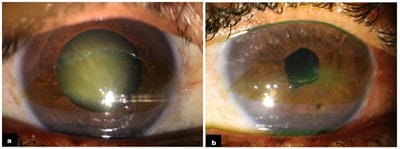
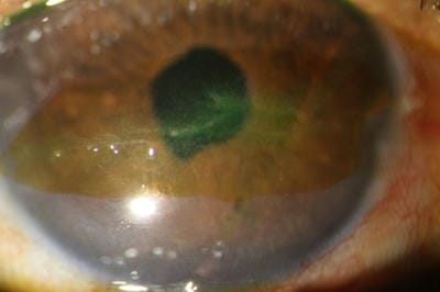
A 54-year-old female presented with complaints of blurring in the right eye since 2 years. A grade 3 nuclear sclerosis and a peculiar band-shaped subepithelial brown corneal patch, within the inter-palpebral area was noted. The overlying epithelium was scraped for smears and cultures which came back negative. The patient underwent uneventful cataract surgery, but returned 2 weeks later with complaints of watering and grittiness. Surprisingly a dendritic ulcer was noted within the pigmented patch, which responded to topical antivirals and tear substitutes. History of a similar episode, 3 years back was elicited upon questioning the patient.

Keywords: HSV keratitis, Corneal pigmentation, Subepithelial plaque

Corresponding Author How to Cite this Article To Browse
Amrapali Bobade Veer, (MBBS, DOMS, DNB), Department of Cornea, PBMA’s H V Desai Eye Hospital, Pune, Maharashtra, India.
Email:
Joshi H, Veer AB, Joshi S, Deshpande M. Brown’y points for guessing!- A case report of an unusual pigmented corneal plaque.. Trop J Ophthalmol Otolaryngol. 2021;6(3):46-49.
Available From
https://opthalmology.medresearch.in/index.php/jooo/article/view/200

Manuscript Received Review Round 1 Review Round 2 Review Round 3 Accepted
2021-06-01 2021-06-10 2021-06-16 2021-06-20 2021-06-25
Conflict of Interest Funding Ethical Approval Plagiarism X-checker Note
No Nil Yes 7%

© 2021 by Hema Joshi, Amrapali Bobade Veer, Shilpa Joshi, Madan Deshpande and Published by Siddharth Health Research and Social Welfare Society. This is an Open Access article licensed under a Creative Commons Attribution 4.0 International License https://creativecommons.org/licenses/by/4.0/ unported [CC BY 4.0].

Introduction

Herpes simplex virus (HSV) is a DNA virus commonly affecting the mouth, genitals and eyes in humans. HSV keratitis is a leading cause of corneal blindness in the developed countries.[1]. Clinically the commonest form encountered is ‘Epithelial Keratitis’ which may present as simple superficial punctate erosion (SPE’s), dendritic ulcer, or larger geographic ulcer [2]. Diagnosis is often clinical and treatment is simple. The virus however, is notorious to establish latency and recur even after successful treatment with antiviral agents. [3,4]. Also in some cases the presentation may be atypical, delaying the diagnosis and appropriate treatment, and may lead to visual morbidity. In this case we present a peculiar, brown-pigmented subepithelial corneal plaque, in an otherwise quiet eye, which developed a superimposed dendritic ulcer within the pigmented plaque, following uneventful cataract surgery.

Case description

A 54-year-old housewife presented to our Cornea Clinic with chief complaints of diminution of vision in the right eye for 2 years. The best-corrected Visual acuity (VA) was Finger Counting at 2 metres. There was no history of trauma, previous ocular surgery, or systemic disorders.

The slit-lamp examination revealed an unusual, brown coloured, band-shaped plaque in the interpalpebral cornea, extending horizontally from limbus to limbus, measuring 12mm x 5 mm, with well-defined margins and a smooth surface. [figure 1a] The plaque was subepithelial, with an intact overlying epithelium, as was noted after the initial scraping.

A grade 3 nuclear sclerosis cataract was also noted in the right eye.

The remaining anterior and posterior segment evaluation was within normal limits.

The lesion was scraped with a 15 no. blade for microbiological assessment. KOH mount, Gram stain, and Giemsa stain came back negative for fungus and bacteria. Blood agar and Sabouraud dextrose agar showed no growth at end of 2 weeks. PCR could not be done due to financial constraints.

As the eye was unremarkable except for the pigmented plaque, the patient was advised cataract surgery for that eye. The surgery was uneventful.

At the 1-week postoperative visit, uncorrected VA in the right eye was 6/36, the eye was quiet, and anterior and posterior segment findings were within normal limits. Steroids were tapered and the patient was given a follow-up date after 3 weeks.

However, the patient returned after a week with complaints of watering, grittiness, and redness. Uncorrected VA had dropped by one line, to 6/60, and slit-lamp examination showed the presence of a dendriform patterned corneal ulcer, within the pigmented plaque. [figure 1b] Well stained with 2% fluorescein and Rose Bengal dye. [figure 2]

Upon questioning again, the patient recalled a similar episode in the right eye, 3 years ago with similar symptoms. The patient has visited a local doctor who gave her topical medication (records unavailable). The symptoms had persisted for a few days and were resolved following treatment.

Clinical diagnosis of HSV keratitis was made and the patient was put on a course of topical acyclovir 3% eye ointment 5 times/day and tear substitutes. Topical steroids were withheld. The symptoms resolved over 2 weeks and the final visual acuity at 1 month was 6/18. The pigmented plaque persisted.

Discussion

Herpes simplex virus (HSV) is a DNA virus that commonly affects the mouth, genitalia, and eye. Ocular involvement is in the form of HSV keratitis, a leading cause of corneal blindness in developed countries.[1]. Clinically HSV keratitis can be classified into epithelial, stromal, and endothelial types. The epithelial keratitis may present as simple corneal erosions, dendritic ulcers, geographic ulcers, or marginal ulcers.[2].

Although easy to treat, HSV keratitis is notorious to establish a latent infection and cause reactivation. [3,4]. Hence complete eradication of the disease is not possible even with antiviral drugs. During active infection, the virus replicates and the virions travel to trigeminal ganglion and establish latency. From time to time, either spontaneously or due to any stressors, the virions travel from the ganglion back to the eye, thus causing a recurrent disease.

Dendritic lesions are pathognomic of HSV keratitis, and diagnosis is primarily clinical but an atypical presentation may impede accurate diagnosis and thus delay appropriate primary treatment and increase the recurrence rates.


In this particular case, a literature review done to find an etiology for the peculiar pigmented plaque did not yield any tangible results. A working hypothesis of iron/ferritin pigment deposition due to the accumulation of tear-film over an irregular ocular surface was made.

Iron is present extracellularly in the tear film on the surface of the cornea. It is carried by lactoferrin which is secreted by acinar epithelial cells in the lacrimal gland. In the tears, more than 90% of lactoferrin is not bound to iron and is available as a chelator and performs anti-oxidative and anti-microbial functions. [5,6].

Excess iron deposition leading to brownish corneal iron lines is observed in many conditions such as Fleischer's Ring in keratoconus, Stocker's Line in a pterygium, Ferry's Line anterior to a filtering trabeculectomy bleb, and the Hudson Stahli line seen as an age-related iron deposition.

Various hypotheses have been put forth to explain the pathophysiology of iron deposition.[7]. These include; Tear Pooling Hypothesis (Gass 1964), Basal-Cell-Migration theory (Rose and Lavin 1987), Tear Desiccation hypothesis (Assil 1993), Senescent basal cell hypothesis (Assil in 1993).

Assil proposed that iron deposition occurs in the areas of tear desiccation, rather than in areas of pooling, and evaporation of the tears increases the relative concentration of iron.[8]. Iron lines typically exist in the basal epithelial cells of the cornea (similar in location to the pigmented plaque seen in our case), and electron microscopy of these lines, demonstrates abundant ferritin.[9].

opthal_200_01.jpg

Figure 1: (a) Brown pigmented corneal subepithelial plaque with intact overlying epithelium noted in the interpalpebral area, in a phakic eye at first visit. (b) Dendritiform ulcer developed 2 weeks after an uneventful cataract surgery that responded to topical antivirals.

Figure 2: Dendritic ulcer staining with 2% fluorescein and rose Bengal dye

opthal_200_02.jpg

Conclusion

A similar past episode recounted by the patient may have been a mistreated epithelial HSV ulcer. Local rural practitioners are known to prescribe topical antibiotics + steroid combinations in cases of corneal ulcers. The resultant conversion of a dendritic ulcer to a geographic ulcer is a common occurrence in such cases. A long-standing, non-healing, geographic ulcer may cause pooling and desiccation of tears, subsequently leading to deposition of iron. This is conjecture, as we have not found any similar cases documented in the literature.

The authors certify that they have obtained all appropriate patient consent forms. The patient has consented to his clinical information and photographs being reported in a scientific journal. The patient understands that his name will not be published and due efforts will be made to conceal the identity, but anonymity cannot be guaranteed.

Reference

01. Darougar S, Wishart MS, Viswalingam ND. Epidemiological and clinical features of primary herpes simplex virus ocular infection. Br J Ophthalmol. 1985 Jan;69(1)2-6. doi: 10.1136/bjo.69.1.2 [Crossref][PubMed][Google Scholar]

02. Holland EJ, Schwartz GS. Classification of herpes simplex virus keratitis. Cornea. 1999 Mar;18(2)144-54. doi: 10.1097/00003226-199903000-00002 [Crossref][PubMed][Google Scholar]


03. Toma HS, Murina AT, Areaux RG Jr, Neumann DM, Bhattacharjee PS, Foster TP, Kaufman HE, Hill JM. Ocular HSV-1 latency, reactivation and recurrent disease. Semin Ophthalmol. 2008 Jul-Aug;23(4)249-73. doi: 10.1080/08820530802111085 [Crossref][PubMed][Google Scholar]

04. Perng GC, Jones C. Towards an understanding of the herpes simplex virus type 1 latency-reactivation cycle. Interdiscip Perspect Infect Dis. 2010;262415. doi: 10.1155/2010/262415 [Crossref][PubMed][Google Scholar]

05. Shimmura S, Suematsu M, Shimoyama M, Tsubota K, Oguchi Y, Ishimura Y. Subthreshold UV radiation-induced peroxide formation in cultured corneal epithelial cells- the protective effects of lactoferrin. Exp Eye Res. 1996 Nov;63(5)519-26. doi: 10.1006/exer.1996.0142 [Crossref][PubMed][Google Scholar]

06. Ellison RT 3rd. The effects of lactoferrin on gram-negative bacteria. Adv Exp Med Biol. 1994;357;71-90. doi: 10.1007/978-1-4615-2548-6_8 [Crossref][PubMed][Google Scholar]

07. Loh A, Hadziahmetovic M, Dunaief JL. Iron homeostasis and eye disease. Biochim Biophys Acta. 2009 Jul;1790(7)637-49. doi: 10.1016/j.bbagen.2008.11.001 [Crossref][PubMed][Google Scholar]

08. Assil KK, Quantock AJ, Barrett AM, Schanzlin DJ. Corneal iron lines associated with the intrastromal corneal ring. Am J Ophthalmol. 1993 Sep 15;116(3)350-6. doi: 10.1016/s0002-9394(14)71353-4 [Crossref][PubMed][Google Scholar]

09. Hiratsuka Y, Nakayasu K, Kanai A. Secondary keratoconus with corneal epithelial iron ring similar to Fleischer's ring. Jpn J Ophthalmol. 2000 Jul-Aug;44(4)381-6. doi: 10.1016/s0021-5155(00)00179-9 [Crossref][PubMed][Google Scholar]