E-ISSN:2456-6454
P-ISSN:2581-4907
RNI:MPENG/2017/74152

Research Article

Fistula

Tropical Journal of Ophthalmology and Otolaryngology

2021 Volume 6 Number 3 May-June
Publisherwww.medresearch.in

Ocular Manifestations Of Carotid Cavernous Fistula and Clinical Outcome After Management

Gantela S.1*, Chowdary N.2, M. S.3
DOI: https://doi.org/10.17511/jooo.2021.i03.01

1* Sirisha Gantela, MS Ophthalmology Professor, Department of Ophthalmology, NRI Medical College and General Hospital, Guntur, Andhra Pradesh, India.

2 N. Lakshmi Chowdary, MS Ophthalmology Professor, Department of Ophthalmology, NRI Medical College and General Hospital, Guntur, Andhra Pradesh, India.

3 Satyanarayana M., MD DM Neuroradiology Interventional radiologist Associate professor, Department of Radiology, NRI Medical College and General Hospital, Guntur, Andhra Pradesh, India.

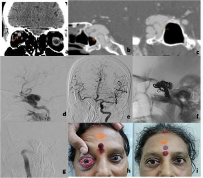
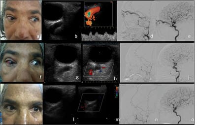
Aim: The purpose of the study is to report the ocular manifestations of Carotid cavernous fistula. To confirm the diagnosis by radiological investigations and to evaluate the clinical outcome after management. Materials and Methods: Patients who presented to the ophthalmology department with signs and symptoms of carotid-cavernous fistula were evaluated by clinical examination followed by radiological investigations like ultrasound, Doppler, CT scan and MRI. They later underwent DSA ( Digital Subtraction Angiography) for confirmation followed by definitive treatment. Results: Out of four patients who had direct CCF, three cases were managed by endovascular embolization of the parent artery. The remaining one case was conservatively managed by carotid massage as it was a low flow fistula and the patient also had Parkinson’s disease. Four cases that had indirect CCF were managed by carotid massage. Complete closure of the fistula is seen in all cases. Patients were followed up for 1 month, 3 months and 6 months and clinical outcome was evaluated. Conclusion: CCF should be suspected in the presence of arteriolised conjunctival vessels, proptosis and audible bruit. Diagnosis is by radiological tests like ultrasonography, Doppler, CT scan and MRI. The confirmatory test is digital subtraction angiography (DSA). Direct CCF is effectively treated with endovascular therapy by coiling the fistula and indirect CCF is managed by manual compression. Early diagnosis and treatment can prevent sight-threatening complications.

Keywords: Carotid Cavernous Fistula, Digital Subtraction Angiography, Endovascular management, Manual Carotid Compression, Proptosis

Corresponding Author How to Cite this Article To Browse
Sirisha Gantela, MS Ophthalmology Professor, Department of Ophthalmology, NRI Medical College and General Hospital, Guntur, Andhra Pradesh, India.
Email:
Gantela S, Chowdary NL, Satyanarayana M. Ocular Manifestations Of Carotid Cavernous Fistula and Clinical Outcome After Management. Trop J Ophthalmol Otolaryngol. 2021;6(3):40-45.
Available From
https://opthalmology.medresearch.in/index.php/jooo/article/view/199

Manuscript Received Review Round 1 Review Round 2 Review Round 3 Accepted
2021-05-04 2021-05-14 2021-05-26 2021-06-05 2021-06-12
Conflict of Interest Funding Ethical Approval Plagiarism X-checker Note
No Nil Yes 8%

© 2021 by Sirisha Gantela, N. Lakshmi Chowdary, Satyanarayana M. and Published by Siddharth Health Research and Social Welfare Society. This is an Open Access article licensed under a Creative Commons Attribution 4.0 International License https://creativecommons.org/licenses/by/4.0/ unported [CC BY 4.0].

Introduction

Carotid Cavernous Fistula (CCF) is the abnormal communication between the carotid artery and the cavernous sinus. The most common cause of CCF is trauma from a basal skull fracture resulting in a tear in the internal carotid artery within the cavernous sinus [1]. Barrow has classified CCF into four types. Type A is the high flow shunt between the internal carotid artery and the cavernous sinus. Types B, C and D are the dural shunts between meningeal branches of the internal carotid and external carotid arteries with the cavernous sinus [2, 3, 4].

The symptoms of CCF are pulsatile proptosis, redness, lid swelling, defective vision, diplopia, ptosis, headache and intolerable bruit. Bruit can be heard using the bell of the stethoscope over the closed eye [5]. The classical signs of CCF are pulsatile proptosis, chemosis and bruit which together are called Dandy’s triad. Conjunctival congestion can be mistaken for chronic conjunctivitis or episcleritis [5,6]. Complications like exposure keratopathy, persistent diplopia, retinal vessel occlusions and secondary glaucoma should also be treated [7,8, 9].

Computerized Doppler imaging, orbital ultrasonography, contrast-enhanced CT scan and MRI are done to diagnose CCF. A confirmatory test for definitive diagnosis is DSA to analyse the venous drainage patterns. Enlargement of the superior ophthalmic vein(hockey stick sign) is a diagnostic feature of CCF (5)

Direct fistulas can be managed with either occluding the fistulous communication or parent artery occlusion, depending on the cross circulation and the number of fistulous communications. The approach can be either transarterial or transvenous. Materials commonly used are balloons, coils and onyx [3,10,11,13]. Few cases can have residual diplopia and visual loss due to compressive optic neuropathy, secondary glaucoma and retinopathy [12]. Most of the patients with dural shunts improve spontaneously or after diagnostic angiography.

Conservative management with carotid artery compression was effective in indirect CCF cases [3, 10, 11, 13].

Materials and Methodology

Eight cases of CCF presenting with ocular manifestations were analysed. Four cases were indirect type and four cases were direct type. Patients were referred from Neurology, Neuro Surgery departments, Interventional radiologist and cases coming to the Ophthalmology department. After taking detailed history complete Ophthalmological examination was done including a Slit-lamp examination, fundus examination with 90 D lens, visual acuity with Snellen’s chart, colour vision with Ishihara chart, Extra Ocular Movements, non-contact tonometry, examination of proptosis and exophthalmometry with Ludde’s exophthalmometer.

Radiological investigations like CT scan, MRI, angiogram, Ultrasound Doppler and DSA were performed. Direct fistulas were managed by the occlusion of the abnormal communication by transvenous or transarterial approach by using coils. Out of eight cases four cases had direct CCF three of these cases were managed by endovascular coiling. One case had Parkinson’s disease also and had low-grade flow in the superior ophthalmic artery and had no cortical venous reflux, this patient improved with manual carotid compression. Four cases with Indirect CCF cases were managed by manual carotid compression. The patients were instructed to compress the lesion side carotid artery with fingers for 10 seconds each time for 15 minutes three times a day. Cases were followed up for 1 month, 3 months and 6 months. Clinical outcome and regression of chemosis or proptosis were observed.

Duration of the study: 3 Years (Jan, 2018 to Jan, 2021)

Ethical Considerations: The study was approved by the institutional ethics committee.


Results

Table No 1 Case details.

Case No Age (yrs) / Sex Etiology Type of Fistula Treatment
1 50 / F Spontaneous Indirect Manual carotid compression
2 60 / F Spontaneous Indirect Manual carotid compression
3 45 / F Trauma RTA fall from the bike Direct Coiling and embolization of parent artery
4 27 / M Trauma RTA post craniotomy for SDH Direct Coiling and embolization of parent artery

5 45 / F Orbitial Rhino Cerebral Mucormycosis Direct Coiling and embolization of parent artery
6 52 / M RTA Indirect Manual carotid compression
7 69 / F RTA Direct Manual carotid compression
8 45/M Spontaneous Indirect Manual carotid compression

Table No 2 Signs in CCF cases before treatment are described in Table No 2

Signs & Symptoms Case 1 Indirect CCF Case 2 Indirect CCF Case 3 Direct CCF Case 4 Direct CCF Case 5 Direct CCF Case 6 Indirect CCF Case 7 Direct CCF Case 8 Indirect CCF
Visual Acuity 6/18 6/24 6/60 6/36 NO PL 6/24 PL+ 6/18
Proptosis 16mm 18mm 18mm 25mm 22mm 26mm 20mm 18mm
Extraocular Movements restriction + + + + + + + +
Fundus Examination Normal fundus Normal fundus Superficial, dot and blot haemorrhages Venous engorgement of vessels CRAO Normal fundus Normal fundus Normal fundus
 IOP 24 mmHg 20.6 mmHg 24.4 mmHg 23.1 mmHg 22 mmHg 18 mmHg 20.6 mmHg 14 mmHg

Table No 3 Signs in CCF cases after treatment are described in Table No 3

Signs & Symptoms Case 1 Indirect CCF Case 2 Indirect CCF Case 3 Direct CCF Case 4 Direct CCF Case 5 Direct CCF Case 6 Indirect CCF Case 7 Direct CCF Case 8 Indirect CCF
Visual Acuity 6/9 6/12 6/18 6/18 NO PL 6/12 6/36 6/12
Proptosis 10mm 14mm 14mm 18mm 16mm 18mm 16mm 15mm
Extra ocular Movements Full and free Full and free Mild Restriction + Full and free Mild Restriction + Full and free Full and free Full and free
IOP 14 mmHg 18 mmHg 17 mmHg 17 mmHg 20 mmHg 18 mmHg 18 mmHg 12 mmHg

Visual acuity improved gradually in all the cases except in one case in which there was a complication of CRAO(Central retinal artery obstruction). High Intraocular pressure was noted in four cases it was treated with suitable antiglaucoma drugs and these drugs were discontinued after one month after the intraocular pressure became normal. Signs like chemosis, congestion, ophthalmoplegia and proptosis were resolved after the treatment in all the cases.

opthal_199_01.jpg

Case Reports: Case Details of Direct CCF are

shown in figure no 1.

One case had a transient increase in the congestion and chemosis immediately after the treatment due to cavernous sinus thrombosis which improved in one month follow up. Review scans were done in all the cases to see for complete closure of the fistula.

Fig 1: Direct CCF. A: Plain CT shows dilated right superior ophthalmic vein (arrow). B & C: CT angiography shows multiple fistulas in the cavernous segment of the right ICA (arrows). D: Right ICA angiogram shows direct carotico cavernous fistula with venous drainage anteriorly into a superior ophthalmic vein, superiorly into cortical veins and inferiorly into basilar plexus. E: Balloon occlusion test revealed good crossflow. F: Coiling of the sac and the parent artery. G: Complete occlusion of right ICA. H & I: Pre and post-op images of the patient.

Fig 2: Indirect CCF. A: Preoperative image of the patient. B & C: USG and doppler of right orbit reveal dilated SOV with the arterialized flow. D & E: right ECA and ICA angiogram revealed fistulous communication with right cavernous sinus with slow flow anteriorly into SOV. No evidence of cortical venous reflux.


opthal_199_02.jpg

Case Details of Indirect CCF are shown in figure no 2.

She was advised carotid compression because of no progressive vision loss, No cortical venous reflux, Low flow shunt, Only anterior superior ophthalmic vein is seen in drainage and No neurological symptoms and signs.

One month later she presented with an increase in redness with pain in the right eye. USG and doppler of right orbit reveal dilated SOV with no flow & right ECA angiogram reveals no evidence of fistula. Right ICA angiogram revealed few communications with the right cavernous sinus.

She was administered inj. Heparin and was advised to continue carotid compression.

On follow up after one month, there was no chemosis or proptosis and the doppler of right orbit reveals a decrease in size of SOV with no flow. The right ECA and ICA angiogram revealed no evidence of fistulous communication.

Discussion

Out of eight cases of CCF, four cases were direct CCf and four cases were indirect CCF. All the cases had ocular manifestations like proptosis, chemosis, arterialization of conjunctival blood vessels seen as cock screw vessels and restriction of ocular movements. Complete ophthalmological examination including visual acuity with Snellen’s chart, colour vision with Ishihara charts, slit lamp examination, fundus examination using 90 D lens, Non-contact tonometry and exophthalmometry were done.

Patients were evaluated before and after the management. Out of four patients who had direct CCF three cases were managed by endovascular embolization of the parent artery.

The remaining one case was conservatively managed by manual carotid massage as it was a low flow fistula and the patient also had Parkinson's disease.

Four cases that had indirect CCF were managed by manual carotid massage. One patient had transient worsening of clinical features like proptosis and chemosis due to cavernous sinus thrombosis. These features resolved gradually and the patient improved in two months duration. Complete closure of fistula is seen in all the cases

In our study the most common cause for CCF is trauma which is comparable to the study done by Miller et al [1]. Four out of eight cases had a history of trauma, head injuries in road traffic accidents, three cases that had spontaneous indirect CCF and one case developed CCF after surgery for orbital rhino cerebral mucormycosis. One case developed CCF after craniotomy was done for subdural haemorrhage following head injury in a road traffic accident.

Raised IOP is seen in all most all cases in the present study. Similar results were described in the study done by Ishijima et al [8] in 43 cases of CCF over 16 years. The IOP was controlled after treatment.

The ocular manifestations like pulsatile proptosis, chemosis, Cock screw vessels and restricted ocular movements were seen in all cases in the present study. One case had complete loss of vision due to central retinal artery occlusion. Poor outcome was due to complications of CCF. These are similar to the findings described by Jayanti Das et al, [4] Imtiaz A Chowdhary [6] and Anna CS Tan [12].

A study was conducted by Anna CS Tan and Sadiaa Farooqui [12] in 45 CCF cases in Asian patients, concluded that clinical features are related to the angiography findings but outcomes may not always correlate after endovascular treatment. Three cases with direct CCF were treated successfully with endovascular management by embolization and coiling in the present study. These cases had improvement in visual acuity and other signs like proptosis, chemosis and congestion were reduced. These results are comparable with that of the results obtained from studies done by Bin du et al [13] and Prechawat et al [10] who studied 80 cases of CCF.

Cases with indirect CCF were well managed by manual compression.


Similar results were described in studies done by Prechawat et al, [10] Adam I Lewis [11] and Bin Du et al [13].

The limitation of this study is the small sample size as CCF is a very rare condition.

Conclusion

Ophthalmologists should work together as a team with neurologists, neurosurgeons and interventional radiologists. CCF should be suspected by observing clinical features like arterialized conjunctival vessels or cock screw vessels, pulsatile proptosis and audible bruit. The investigation of choice for confirmation of CCF is digital subtraction angiography. Direct CCF is effectively treated by embolization with endovascular therapy and indirect CCF is managed by manual compression. Early diagnosis and management can prevent sight-threatening complications.

Contribution Details

Dr.Gantela Sirisha has conceptualized the study prepared the study protocol, conducted the Ophthalmological examination, data collection and manuscript writing. Dr.Gantela Sirisha has verified all the drafts and approved the final draft. Dr. M. Satyanarayana has done radiological diagnosis and performed endovascular embolization procedures. Dr. N. Lakshmi Chowdary has provided key inputs and supported the study. Dr. M. Satyanarayana, Dr. N. Lakshmi Chowdary has also edited all the drafts and approved the final draft of the manuscript.

Abbreviations

CCF: Carotid Cavernous Fistula

DSA: Digital Subtraction Angiogram

SOV: Superior Ophthalmic Vein

Reference

01. Miller, Neil R. "Diagnosis and management of dural carotid–cavernous sinus fistulas". Neurosurgical Focus. 23;5(2007)E13. [Crossref][PubMed][Google Scholar]

02. Barrow DL, Spector RH, Braun IF, Landman JA, Tindall SC, Tindall GT. Classification and treatment of spontaneous carotid-cavernous sinus fistulas. J Neurosurg. 1985;62(2)248-56. doi: 10.3171/jns.19 85.62.2.0248 [Crossref][PubMed][Google Scholar]

03. Chi CT, Nguyen D, Duc VT, Chau HH, Son VT. Direct traumatic carotid cavernous fistula- angiographic classification and treatment strategies, Study of 172 cases. Interv Neuroradiol. 2014 Jul-Aug;20(4)461-75. doi: 10.15274/INR-2014-10020 [Crossref][PubMed][Google Scholar]

04. Das JK, Medhi J, Bhattacharya P, Borah N, Bhattacharjee K, Kuri GC, et al. Clinical spectrum of spontaneous carotid-cavernous fistula. Indian J Ophthalmol. 2007 Jul-Aug;55(4)310-2. doi: 10.4103/0301-4738.33051 [Crossref][PubMed][Google Scholar]

05. Myron Yanoff & Jays S Duker. Text Book of Ophthalmology, 4th Edition. Elsevier Part 9 Neuro Ophthalmology Section 5 Neuro Ophthalmologic Emergencies & Vascular Disorders. Page No- 993-995. [Crossref][PubMed][Google Scholar]

06. Chaudhry IA, Elkhamry SM, Al-Rashed W, Bosley TM. Carotid cavernous fistula- ophthalmological implications. Middle East Afr J Ophthalmol. 2009 Apr;16(2)57-63. doi: 10.4103/0974-9233.53862 [Crossref][PubMed][Google Scholar]

07. Bhatti MT, Peters KR. A red eye and then a really red eye. Surv Ophthalmol. 2003 Mar-Apr;48(2)224-9. doi: 10.1016/s0039-6257(02)00461-7 [Crossref][PubMed][Google Scholar]

08. Ishijima K, Kashiwagi K, Nakano K, Shibuya T, Tsumura T, Tsukahara S. Ocular manifestations and prognosis of secondary glaucoma in patients with carotid-cavernous fistula. Jpn J Ophthalmol. 2003 Nov-Dec;47(6)603-8. doi: 10.1016/j.jjo.2003.08.002 [Crossref][PubMed][Google Scholar]

09. de Keizer R. Carotid-cavernous and orbital arteriovenous fistulas- ocular features, diagnostic and hemodynamic considerations in relation to visual impairment and morbidity. Orbit. 2003 Jun;22(2)121-42. doi: 10.1076/orbi.22.2.121.14315 [Crossref][PubMed][Google Scholar]

10. Preechawat P, Narmkerd P, Jiarakongmun P, Poonyathalang A, Pongpech SM. Dural carotid cavernous sinus fistula- ocular characteristics, endovascular management and clinical outcome. J Med Assoc Thai. 2008 Jun;91(6)852-8. [Crossref][PubMed][Google Scholar]


11. Lewis AI, Tomsick TA, Tew JM Jr. Management of 100 consecutive direct carotid-cavernous fistulas- results of treatment with detachable balloons. Neurosurgery. 1995 Feb;36(2)239-44; discussion 244-5. doi: 10.1227/00006123-199502000-00001 [Crossref][PubMed][Google Scholar]

12. Tan AC, Farooqui S, Li X, Tan YL, Cullen J, Lim W, et al. Ocular manifestations and the clinical course of carotid cavernous sinus fistulas in Asian patients. Orbit. 2014 Feb;33(1)45-51. doi: 10.3109/01676830.2013.851253 [Crossref][PubMed][Google Scholar]

13. Du, Bin, et al. A retrospective analysis of 38 carotid cavernous fistula patients treated with balloon-assisted endovascular fistula embolization through simultaneous transarterial and transvenous approaches". Int J Clin Exp Med. 9;10(2016)19399-19407. [Crossref][PubMed][Google Scholar]