E-ISSN:2456-6454
P-ISSN:2581-4907
RNI:MPENG/2017/74152

Research Article

Cataracts

Tropical Journal of Ophthalmology and Otolaryngology

2020 Volume 5 Number 8 November-December
Publisherwww.medresearch.in

Incidence, management, and outcome of complex cataracts in a tertiary eye set up

Verma R.1, Shrishrimal M.2, Gokharu S.3, Shah D.4*
DOI: https://doi.org/10.17511/jooo.2020.i08.02

1 Ritu Verma, Cornea and Cataract Consultant, Department of Ophthalmology, Choithram Netralaya, Shriram Talawali, Indore, Madhya Pradesh, India.

2 Meghna Shrishrimal, Senior Resident, Department of Ophthalmology, Choithram Netralaya, Shriram Talawali, Indore, Madhya Pradesh, India.

3 Shirali Gokharu, DNB Resident, Department of Ophthalmology, Choithram Netralaya, Shriram Talawali, Indore, Madhya Pradesh, India.

4* Dhaivat Shah, Vitreoretina Consultant, Department of Ophthalmology, Choithram Netralaya, Shriram Talawali, Indore, Madhya Pradesh, India.

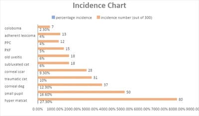
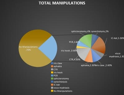
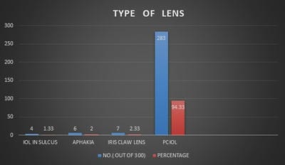
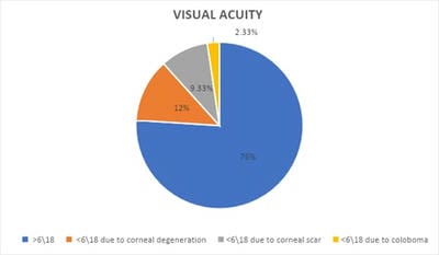
Aim: To study the incidence, management, and outcome of complex cataract cases in a tertiary eye care center. Material and methods: Retrospective observational study with analysis of records done for patients who were operated on for complex cataract from December 2019 to February 2020. Results: 300/10,000 patients had complex cataracts requiring additional surgical skills, advanced equipment, and better patient counseling. 5.3% (16/300) of patients needed intracameral adrenaline whereas 1.3% (4/300) needed Visco mydriasis. A capsular tension ring was implanted in 4.3%(13/300) of patients. 4% (12/300) underwent sphincterotomies whereas 2% (6/300) had to undergo synechiolysis. Iris hooks were used in only 0.6% of patients (2/300). Posterior capsular rent was seen in 1.6%(5/300) which was managed by placing a Posterior chamber IOL in the sulcus. 2.6% (8/300) had implantation of the iris-claw lens and 2.3% (7/300) were left aphakic for secondary IOL to be implanted at a later date. The visual outcome was better than 6/18 in 76% of patients. 24% of patients had an improvement in their visual acuity by a line or 2 but was less than 6/18 due to corneal degenerations, corneal scarring, Retinal pathologies, and colobomas. Conclusion: Although these cataracts demand more expertise on behalf of the surgeon, the outcome can be very rewarding if done with proper planning. The knowledge of small incision cataract surgery is highly valuable in such situations.

Keywords: Complex cataracts, Intraoperative Manipulations, Visual outcome

Corresponding Author How to Cite this Article To Browse
Dhaivat Shah, Vitreoretina Consultant, Department of Ophthalmology, Choithram Netralaya, Shriram Talawali, Indore, Madhya Pradesh, India.
Email:
Verma R, Shrishrimal M, Gokharu S, Shah D. Incidence, management, and outcome of complex cataracts in a tertiary eye set up. Trop J Ophthalmol Otolaryngol. 2020;5(8):207-215.
Available From
https://opthalmology.medresearch.in/index.php/jooo/article/view/174

Manuscript Received Review Round 1 Review Round 2 Review Round 3 Accepted
2020-11-18 2020-11-28 2020-12-10 2020-12-24
Conflict of Interest Funding Ethical Approval Plagiarism X-checker Note
No Nil Yes 6%

© 2020 by Ritu Verma, Meghna Shrishrimal, Shirali Gokharu, Dhaivat Shah and Published by Siddharth Health Research and Social Welfare Society. This is an Open Access article licensed under a Creative Commons Attribution 4.0 International License https://creativecommons.org/licenses/by/4.0/ unported [CC BY 4.0].

Introduction

India has been very successful in increasing its cataract surgical rate and now is at par with the estimate of 8000-8700 per million cataract surgeries to eliminate blindness [1,2]. The reason for this surge in the rate of cataract surgeries is due to increased awareness among the population, increase in the facilities to accommodate more patients in the hospital premises, their transportation, and meals. This number was attained because of the increase in the efficiency of surgeons and the availability of the latest infrastructure in the eye hospitals. The District Blindness Control Societies organizes camps in remote villages that do not have access to hospitals leading to an increase in the number of patients enrolling for cataract surgery in each camp [3]. ​With more patients registering for surgery in a tertiary eye setup, there is also an increase in difficult, complex cataracts which are denied in other smaller private setups with lesser infrastructure. It is a well-known fact that each cataract behaves differently in the operating room. Certain cataracts are at a higher risk of complications during surgery. Surgery on such complex cataracts yields guarded visual outcomes even when they demand more surgical expertise and advanced machinery. Recognizing such complex cataracts helps us to be prepared to deal with the likely complications. These patients need more counseling regarding the guarded visual outcome so that they have a realistic expectation and in turn improve follow-up and medication compliance. It helps the OT personnel to make the necessary equipment ready and available during surgery [4]​.

Complex cataracts involve the following findings:

Corneal Degenerations and Thinning: Corneal degenerations like Climatic droplet keratopathy and band-shaped keratopathy can make the surgery a task due to the hazy visualization. Decreasing the frequent instillation of phenylephrine drops as it increases corneal haziness, increased use of viscoelastic on the corneal surface, enhancing the visualization of the capsule by staining makes the surgical steps easier.

Corneal and scleral thinning poses a problem in wound healing and hence appropriate measures need to be taken during surgery like the use of low strength steroids and wound suturing for better integrity.

These measures also help to lower the chances of infection post-surgery.

Adherent Leucoma: Surgery in adherent leucomasis a challenge as the pupil is irregular and sometimes it does not dilate well. Releasing the iris tissue, using iris hooks can be done to help during the surgery [4]​.

Pupil Irregularities: Small rigid pupils are a common occurrence in old age, adherent leucomas, diabetics, and pseudoexfoliation (PXF) syndrome. Small pupils pose a challenge during each step of the surgery. Various methods to deal with it are:

  1. Viscodilation
  2. Sphincterotomy
  3. Iris hooks
  4. Iris rings

Colobomas: Eyes with congenital coloboma are at greater risk for complications during cataract surgery. Given the abnormal development of the sclera, uvea, and zonules, the structural integrity of the globe may be compromised, increasing the intraoperative risks like damage to the corneal endothelium, Descemet's membrane detachment, posterior capsular dehiscence, vitreous loss, and nucleus drop. The risk increases with the severity of microcornea and grade of nuclear sclerosis [5,6,7]​.

Uveitis: Cataract surgery in a uveitic eye requires various manipulations intraoperatively like membranectomy, synechiolysis, use of iris hooks as and when needed, or a combination of all. Uveitic cataracts are difficult to manage postoperatively due to high chances of inflammation and a toxic anterior segment syndrome [8,9].

Pseudoexfoliation syndrome: Pseudoexfoliation (PXF) is a fibrillar material deposited in the anterior segment of the eye and causes difficulties during Cataract surgery [10]. Eyes with pseudoexfoliation have been associated with complications of zonular dehiscence, posterior capsule rupture (PCR), vitreous loss, and lens dislocation intraoperatively [11].

Subluxated and Dislocated cataracts: Ectopia lentis is the dislocation or displacement of the natural crystalline lens. The lens is defined as luxated (dislocated) when it lies completely outside of the hyaloid fossa, and is subluxated when it is partially displaced but remains within the lens space [12].​ Appropriate surgical management of a subluxated cataract is a daunting task.


While smaller degrees of the subluxation can be treated with capsular tension rings, larger degrees of subluxation and dislocation of the lens needs the removal of the lens with the bag. After anterior vitrectomy, any of the following lenses Anterior chamber lens, Iris claw lens, or scleral fixated Lens can be used [13,14].

Traumatic Cataracts: Traumatic cataracts have to be evaluated very thoroughly and carefully as they can give nasty surprises on the table. Scarred tracks of corneal tears, iris trauma, traumatic mydriasis, rupture of the anterior capsule, rupture of the posterior capsule, subluxation varying from mild to severe can be seen alone or in combination. Scarred corneas hamper the visualization intraoperatively and give refractive surprises postoperatively. Both the surgeon and the patient should be prepared for the results [4].​

Posterior Polar Cataract: Posterior polar cataracts (PPCs) are known to be associated with an abnormal adhesion of the posterior capsule to the polar opacity, or a preexisting weakness of the posterior capsule, both of which predispose the eye to the posterior capsule rupture (PCR) during cataract surgery. PPCs are, therefore, a nightmare for all cataract surgeons, and several strategies are being propagated to reduce the PCR rate in these cataracts [15]​.

Hypermature cataracts: Despite all the efforts taken by the government, there is still a lack of awareness among the rural population leading to the late presentation which results in a high incidence of hypermaturecataracts [16]. Intraoperative complications in hypermature cataracts are related to weak zonules and fibrotic capsules sometimes leading to capsulorhexis extension and/or dislocation of the entire capsular bag. Capsulorhexis with high density viscoelastic and capsular tension rings in cases of zonular weakness helps.

Material and Methods

Case records of 300 patients with Complex cataracts operated in the community department of ChoithramNetralaya, Indore from December 2019 to February 2020 were reviewed retrospectively. The screening was done by trained optometrists in outreach camps. Demographic data, preoperative evaluation, intraoperative notes, and postoperative data were reviewed. Demographic data included name, age, sex, and systemic history.

The preoperative evaluation consisted of visual acuity, sac status, IOP, slit lamp evaluation, fundus examination, keratometry, A-Scan, B-scan, blood pressure, and random blood sugar levels. If the patient has diabetes then fasting and postprandial sugar levels were done. ECG was ordered if there was any indication for the same. All the patients were counseled regarding the complexity of their cataracts and the visual prognosis. Written informed consent was taken. All the patients underwent Manual Small Incision cataract surgery by a team of well-experienced surgeons along with subspeciality backup. Any intraoperative manipulations like synechiolysis, sphincterotomies, use of iris hooks, use of Capsular Tension Ring, use of Anterior Vitrectomy, and the type of lens used were noted. Postoperative Day 1 finding and treatment were noted. 1 week follow up findings were recorded. 6 weeks follow up consisted of refraction and best-corrected visual acuity.

Normal Cataracts were excluded from the study. Statistical analysis was done using a parametric method of univariate analysis. Percentage proportions were analyzed for parameters relevant to the study.

Results

Around 10,000 patients underwent cataract surgery from December 2019 to February 2020. Around 3% patients among these had complex cataracts(300/10,000). Among the 300 complex cataracts, the highest incidence was that of hyper mature cataracts coming up to 27.3% (80/300). In 6.1% of hypermature cataracts, a Capsular Tension Ring (CTR) was used. 3.6% of patients were implanted with an iris-claw lens as the capsular bag was explanted due to instability. Aphakia was reported at 1.2%. Common Postoperative day 1 finding was fibrinous membrane in 8.5% which was treated with oral and topical steroids and cycloplegic-mydriatics.

Corneal edema on the first postoperative day was seen in 16% of patients which was alleviated in the 1st-week follow-up. Primary PCO was another common occurrence in 39% and the patients were counseled for YAG Capsulotomy after 3 months. Out of hypermature cataracts, 2 patients had lens-induced glaucoma. The intraocular pressure was reduced preoperatively with 200cc intravenous Mannitol and tablet Acetazolamide 250mg thrice a day. Once the IOP was reduced the patients were taken up for surgery.


The small pupils accounted for 16.66%(50/300). 58% were mature cataracts and 10% were brown-black cataracts. 32% of them were dilated with intracameral adrenaline. Sphincterotomies were done in 10% of cases. 8% of patients underwent Visco mydriasis. Iris hooks were used in 2%. All the patients improved to the vision of 6/18 or better. Pseudoexfoliation syndrome (PXF) was reported in 5%(15/300). 6.6% of the PXF cases had aphakia to be treated with secondary IOLs. 20% of patients underwent sphincterotomies. The rest of the cases were uneventful. The only non-glaucomatouspseud- oexfoliation syndrome was included in this study.

Traumatic cataracts accounted for 10.33% (31/300). Aphakia to be treated with secondary IOLs was seen in 6.45%of patients. Iris claw was implanted in 6.45%. CTR was used in 16% of patients. 3.2% of the patients required synechiolysis. 1 patient had a spontaneous choroidal detachment intraoperatively and was left aphakic which was treated with oral steroids. The patient was later operated on for secondary IOL. 12.33% of patients had corneal degenerations (37/300). Surgeries were uneventful with a visual improvement of 2-3 lines in all the patients. The lesser visual acuity was due to corneal degeneration. Corneal scarring with thinning was seen in 9.3%(28/300). The surgery per se was uneventful in all the patients. There was a visual improvement in all the patients by a line or 2 on Snellen’s chart. Posterior polar cataracts came to 4%(12/300). Posterior capsular rent (PCR) was seen in 8.3%(1/12), and the IOL was placed in the sulcus because of a good capsulorhexis. 6% of patients had subluxated cataracts(18/300). In 66% of subluxated cataracts, the zonular dehiscence was less than 2 clock hours and hence PMMA IOL support was enough and no extra manipulation was needed. CTR was implanted in 17% of patients. 11% of patients needed an iris-claw lens with anterior vitrectomy.

opthal_174_01.jpg

Fig-1: Incidence chart.

Fig-2: Total manipulations.

opthal_174_02.jpg

Fig-3: Type of Lens.

opthal_174_03.jpg

Fig-4: Visual acuity

opthal_174_04.jpg

Another 6% (18/300) of patients had cataracts complicated with old uveitis. 27.7% of patients with old uveitis underwent synechiolysis. 5.5% of patients needed sphincterotomy and another 5.5% of patients required the use of iris hooks. Postoperative inflammation increased by 27.77% with grade 4 cells and flare. 22% of the patients had fibrinous membrane and 27% of patients had corneal edema. All the patients were treated with a tapering course of oral steroids and cycloplegics.


All the patients had improvement in the 1st week follow up and had a quiet eye at 6 weeks. 4.3%(13/300) of patients had adherent leucoma. In 15.38% of patients, sphincterotomy was done. In 7.7% of patients, the adherent leucoma was released. The rest of the surgery was uneventful for all the patients. 2.3 %(7/300) of patients had coloboma. In 28%(2/7) of patients, CTR was used to support the capsular bag. Another 28%(2/7) were left aphakic. But 42.8%(3/7) of the surgery was uneventful as the zonular weakness was not more than 2 clock hours and the capsular bag support was good. Visual outcome was 3/60 in 14.2%(1/7) as the coloboma was involving the macula. 28%(2/7) had vision 6/60 and the rest of the 4 patients were lost to follow up. More than 50% of patients were lost to follow up at our center due to the remoteness of the areas and lack of connectivity. However, these patients are followed up in the district health centers where the ophthalmic assistants checked for visual acuity. On feedback from those centers, authors noted that visual outcome was better than 6/18 in most of the patients (76%). Patients having other ocular comorbidities like corneal pathologies, retinal pathologies had an improvement in the visual acuity by a line or 2 (24%).

Discussion

Pseudoexfoliation: The frequency of PXF syndrome in the present study (5%) was lower than that in a hospital-based study done by Rajesh et al. In the present study, the mean pupil diameter achieved after maximal mydriasis was significantly less in the PEX group which simulates with the study done by Rajesh et al [18].

Scorolli et al [19] and Praveen et al [20] found that patients with PXF have five times a greater risk of intraoperative complications in cataract surgery compared to normal cases. They further stated that the recognition of this condition is very important before starting surgery on such patients. The visual outcome though was good in the present study as well as other studies mentioned above despite the difficult surgery and with proper precautions.

Hypermature cataracts: Hypermature cataracts are a challenge for almost all ophthalmic surgeons as observed in the present study and mentioned in other studies, as this type of cataract exhibits a fibrous​ anterior capsule, a liquid cortex, an expanding capsular bag, zonular weakness, and a hard nucleus [21].

Techniques like visco-shell technique [22]​and intraocular lens (IOL) scaffold​ technique [23], have achieved satisfactory results in studies done by different authors. The incidence of primary PCO in a study conducted by Rajesh et al is 38% as compared to 39% in the present study [17]​. The visual outcome was satisfactory in ours and all the other studies.

Corneal Degenerations and scarring: It makes the surgery difficult due to hindered visualization. Despite the effort, the visual outcome is poor due to the lack of transparency of the cornea. In a study done in Saudi Arabia, the incidence of corneal scarring is 13% and that of climatic droplet keratopathy is 11% [24]. In the present study, the incidence of corneal scarring is a bit lower accounting​ for 9.3% whereas that of corneal degenerations was 12.3%. In a study done in Kuwait corneal degenerations and scarring accounted for poor visual outcomes in 40% amongst complex cataracts as compared to 21.1% in the present study [25].

Small pupil: It's a well-known fact that in a patient whose pupils fail to dilate are more prone to complications like posterior capsular rupture, retained lens matter, vitreous loss [26].

According to Malyugin et al topical medications augmented with intraocular mydriatic injections appear to be the mainstream providing success in 90%–95% of all cases as is seen in the present study. Iris hooks and Malyugin Ring are the current standard of care for intraoperative mechanical pupil expansion in patients not responding to the pharmacological protocols according to Malyuginetal [27]. In our case, only iris hooks were used for mechanical pupil expansion in 2%, and​ sphincterotomies are done in 8% of cases.​

Posterior Polar cataracts: Cortical- cleaving hydrodissection leads to hydraulic rupture of the posterior capsule and hence it should be avoided in PPC [28,29]. Hence in the present study, all the PPC underwent hydrodelineation as it creates an epinuclear cushion and prevents posterior capsular rupture. Hydrodelineation has been recommended in many other studies done by Masket [30], Hayashi​ , et al​​, [31]​ Allen and Wood [32] ​, and Lee and Lee [33]​. In addition to hydrodelineation, Fine et al​​ [34]​ also advised performing hydro dissection in multiple quadrants injecting tiny quantities of fluid gently, such that the fluid wave is not allowed to spread across the posterior capsule.


The risk of posterior capsular rupture has decreased from 36% [28] to 6-7% [31]​ in the recent era. In our​study, the PCR rates are 8.3%(1/12). PCR rates in PPC are declining due to a better understanding of the anatomy of the PPC and modification in the surgical steps to reduce the same [28].

Uveitis: In the present study, the incidence of uveitic cataracts accounts for 6% as compared to 1.2% in a study done by Chu et al [35]. Due to better control of inflammation pre and postoperatively the visual outcomes have improved from 59% to 82% [36]. According to Kosker et al, the visual outcomes are the best in anterior uveitis with 91.1% to 94.5% achieving visual acuity 6/18 or better which is comparable to the present study [37,38]. A steroid cover pre and post-op in such cases can be an asset to control inflammation.

Traumatic cataracts: Attaining a satisfactory visual acuity in traumatic cataract surgery is unpredictable. Lens was implanted in 90% in the present study as compared to 81.6% in a study by Mehul et al [39] and 78.6% by Synderetal [40]. Visual acuity in our case study was better than 6/18 in 90% of cases as compared to 64.7% by Gradinetal [41], Loncar et al [42] also reported good visual outcomes post-traumatic cataract surgeries.

Subluxated cataracts: In cataract surgery with subluxated cataracts, the zonular dialysis mustn't increase in size. This is achieved by using dispersive viscoelastic, careful capsulorhexis with forceps, cortical cleaving hydrodissection, use of endocapsular support devices.

In a study done by Goel et al 77% of subluxated cataracts managed by small incision cataract surgery were successful without the need for any additional procedure as compared to 66% in the present study [43]. Improved visual acuity was achieved in 95% of cases in comparison with 91.7% by Xiao Ma et al [44]. Similar comparable outcomes were seen in 2 other studies [45,46].

Colobomas: Cataract surgery in colobomas is difficult because there are high chances of extension of zonular dehiscence, vitreous herniation, decentration of IOL, or inability to implant IOL. Nordlund et al reported that out of 7 cases 6 patients had an improvement in visual acuity after cataract surgery, whereas one patient developed retinal detachment [5].

Gurler and colleagues reported the successful use of capsular tension ring in cataract surgery in colobomas. They reported 13 cases and all of them had a significant improvement in visual acuity without major complications [47]. Findings in the present study correlate with the above studies.

Conclusion

Complex cataract behaves differently than the normal ones. Although these complicated cases demand more expertise on the behalf of the surgeon and the outcome is guarded, it is worth taking the effort.

What does the study add to the existing knowledge?

A Cataract surgeon of today's era must possess an armamentarium of a spectrum of pharmacological and surgical strategies along with a splendid understanding of ocular anatomy to deliver better outcomes in adverse conditions. If done with proper planning and counseling, the result makes the patient happy and grateful. Manual small incision cataract surgery is a good option in such cases in a community setup where patient load is high and it is cost-effective and can deliver great results.

Author’s contribution

Dr. Ritu Verma: Concept

Dr. Meghna Shrishrimal: Statistical analysis

Dr. Shirali Gokharu: Study design

Dr. Dhaivat Shah: Manuscript preparation

Acknowledgements

Authors would like to acknowledge our team of doctors, optometrist, Operating room staff, and the statistician for their constant help and efforts. Authors are grateful to our patients for having trust and confidence in us.

Reference

  1. Aravind S, Haripriya A, Sumar Taranum BS. Cataract surgery and intraocular lens manufacturing in India. Curr Opin Ophthalmol. 2008;19(1)60-65.

  1. National Program for Control of Blindness- State wise targets and achievements for various eye diseases during 2012–13. 2008;19(1)60-65.
  2. Gudlavalleti VS Murthy, BK Jain, BR Shamanna, D Subramanyam. Improving cataract services in the Indian context. Community Eye Health. 2014;27(85)4-5.
  3. Gogate P Wood M. Recognising ‘high-risk’ eyes before cataract surgery. Community Eye Health. 2008;21(65)12-14.
  4. Nordlund ML, Sugar A, Moroi SE. Phacoemul sification and intraocular lens placement ineyes with cataract and congenital coloboma- visual acuity and complication. J Cataract Refract Surg. 2000;26(7)1035-1040.
  5. Joon-Hyun Kim, Min-Ho Kang, Sung-Min Kang, Byung-Joo Song. A Modified Iris Repair Technique and Capsular Tension Ring Insertion in a Patient with Coloboma with Cataracts. Korean J Ophthalmol. 2006;20(4)246-249.
  6. Sen AC, Kohli GM, Ashish Mitra, Shubhi Tripathi, Sachin B Shetty, Sonal Gupta. Pars-plana vitre ctomy with phacofragmentation for hyperdense cataracts in eyes with severe microcornea and chorio-retinal coloboma- A novel approach. Indian J Ophthalmol. 2020;68(1)91-98.
  7. Alekseev BN, Sibai SA. Opredelenietermina "oslozhnennyekatarakty" [Definition of the term "complicated cataracts"]. Vestn Oftalmol. 1996;112(4)26-27.
  8. Agrawal R, Murthy S, Ganesh SK, Phaik CS, Sangwan V, Biswas J. Cataract Surgery in Uveitis. Int J Inflam. 2012;548453.
  9. Thevi T, Abas AL. Intraoperative and postoperative complications of cataract surgery in eyes with pseudoexfoliation – An 8-year analysis. Oman J Ophthalmol. 2019;12(3)160-5.
  1. Sangal N, Chen TC. Cataract surgery in pseudoexfoliation syndrome. Semin Ophthalmol. 2014;29(5-6)403-408.
  2. Clarke CC. "Ectopia lentis- a pathologic and clinical study". Arch Ophthalmol. 1939;21(1)124-153.
  3. Kim EJ, Berg JP, Weikert MP, Kong L, Hamill MB, Koch DD et al. Scleral-fixated capsular tension rings and segments for ectopia lentis in children. Am J Ophthalmol. 2014;158(5)899–904.
  4. S Khokhar, N Aron,N Yadav, G Pillay, and E Agarwal. Modified technique of endocapsular lens aspiration for severely subluxated lenses. Eye (Lond). 2018;32(1)128-135.
  5. Vasavada AR, Vasavada AV. Managing the posterior polar cataract- An update. Indian J Ophthalmol. 2017;65(12)1350-1358.
  6. Khanna R, Pujari S, Sangwan V. Cataract surgery in developing countries. Curr Opin Ophthalmol. 2011;22(1)10-14.
  7. Joshi RS. Primary posterior capsular opacification in Indian rural population under going cataract surgery for hypermature senile cataract. Clin Ophthalmol. 2013;7;1605-8.
  8. Joshi RS, Singanwad SV. Frequency and surgical difficulties associated with pseudoexfoliation syndrome among Indian rural population scheduled for cataract surgery- Hospital-based data. Indian J Ophthalmol. 2019;67(2)221-226.
  9. Scorolli D, Campos EC, Bassein L, Meduri RA. Pseudoexfoliation syndrome- A cohort study on intraoperative complications in cataract surgery. Ophthalmologica. 1998;212(4)278-280.

  1. Sastry PV, Singal AK. Cataract surgery outcome in patients with non-glaucomatous pseudoexfoliation. Rom J Ophthalmol. 2017;61(3)196-201.
  2. Hua X, Dong Y, Wang L, Li Z, Du J, Chi W, et al. Intraocular lens implantation performed first to protect the posterior capsule in Morgagnian cataracts during phacoemulsification. Int J Ophthalmol. 2019;12(7)1215-1218.
  3. Sato M, Mizushima Y, Oshika T. Visco-shell technique with sodium hyaluronate 2,3% in phacoemulsification of Morgagnian cataract. J Cataract Refract Surg. 2008;34(11)1824-1827.
  4. Parkash RO, Mahajan S, Parkash TO, Nayak V. Intraocular lens scaffold technique to prevent posterior capsule rupture in cases of Morgagnian cataract. J Cataract Refract Surg. 2017;43(1)8–11.
  5. M F al Faran. Visual outcome and complications after cataract extraction in Saudi Arabia. Br J Ophthalmol. 1990;74(3)141-143.
  6. Salem M AL, Ismail L. Factors influencing visual outcome after cataract extraction among Arabs in Kuwait. Br J Ophthalmol. 1987;71(6)458-461.
  7. Bartlett JD, Miller KM. Phacoemulsification techniques for patients with small pupils. Compr Ophthalmol Update. 2003;4;171-176.
  8. Malyugin B. Cataract surgery in small pupils. Indian J Ophthalmol. 2017;65(12)1323-1328.
  9. Vasavada AR, Singh R. Phacoemulsification with posterior polar cataract. J Cataract Refract Surg. 1999;25(2)238-245.
  1. Osher RH, Yu BC, Koch DD. Posterior polar cataracts- a predisposition to intraoperative posterior capsular rupture. J Cataract Refract Surg. 1990;16(2)157-162.
  2. Masket S. Consultation Section. J Cataract Refract Surg. 1997;23(10)819–824.
  3. Hayashi K, Hayashi H, Nakao F, Hayashi F. Outcomes of surgery for posterior polar cataract. J Cataract Refract Surg. 2003;29(1)45-49.
  4. Allen D, Wood C. Minimizing risk to the capsule during surgery for posterior polar cataract. J Cataract Refract Surg. 2002;28(5)742-744.
  5. Lee MW, Lee YC. Phacoemulsification of posterior polar cataracts- a surgical challenge. Br J Ophthalmol. 2003;87(11)1426-1427.
  6. Fine IH, Packer M, Hoffman RS. Management of posterior polar cataract. J Cataract Refract Surg. 2003;29(1)16-19.
  7. Chu CJ, Dick AD, Johnston RL, Yang YC, Denniston AK. UK Pseudophakic Macular Edema Study Group, Cataract surgery in uveitis- A multicentre database study. Br J Ophthalmol. 2017;101(8)1132-1137.
  8. Liu X, Zhao C, Xu T, Gao F, Wen X, Wang M, et al. Visual prognosis and associated factors of phacoemulsification and intraocular lens implantation in different uveitis entities in Han Chinese. Ocul Immunol Inflamm. 2017;25(3)349-355.
  9. Kosker M, Sungur G, Celik T, Unlu N, Simsek S. Phacoemulsification with intraocularlens implantation in patients with anterior uveitis. J Cataract Refract Surg. 2013;39(7)1002-1007.

  1. Chan NSC, Seng-EiTi, Soon-Phaik Chee. Decision-making and management of uveitic cataract. Indian J Ophthalmol. 2017;65(12)1329-1339.
  2. Shah M, Shah S, Shah S, Prasad V, Parikh A. Visual recovery and predictors of visual prognosis after managing traumatic cataracts in 555 patients. Indian J Ophthalmol. 2011;59(3)217-222.
  3. Synder A, Kobielska D, Omulecki W. Intraocular lens implantation in traumatic cataract. Klin Oczna. 1999;101(5)343-346.
  4. Gradin D, Yorston D. Intraocular lens implantation for traumatic cataract in children in East Africa. J Cataract Refract Surg. 2001;27(12)2017-2025.
  5. LacmanovicLoncar V, Petric I. Surgical treatment, clinical outcomes, and complications of traumatic cataract- Retrospective study. Croat Med J. 2004;45(3)310-313.
  6. Goel R, Kamal S, Kumar S, Kishore J, Malik KPS, Bodh SA, et al. Feasibility and Complications between Phacoemulsification and Manual Small Incision Surgery in Subluxated Cataract. J Ophthalmol. 2012;205139.
  1. Xiao Ma, Zhaohui Li. Capsular tension ring implantation after lens extraction for management of subluxated cataracts. Int J Clin Exp Pathol. 2014;Jun 15;7(7)3733-3738.
  2. Jacob S, Agarwal A, Agarwal A, Agarwal S, Patel N, Lal V. Efficacy of a capsular tension ring for phacoemulsification in eyes with zonular dialysis. J Cataract Refract Surg. 2003;(2)315-321.
  3. Georgopoulos GT, Papaconstantinou D, Georgalas I, Koutsandrea CN, Margetis I, Moschos MM. Management of large traumatic zonular dialysis with phacoemulsification and IOL implantation using the capsular tension ring. Acta Ophthalmol Scand. 2007;85(6)653-657.
  4. Gurler B, Coskun E, Okumus S1, Pinero DP, Ozcan E1, Erbagci I. Surgical outcomes of isolated lens coloboma with or without cataract among young adults. Can J Ophthalmol. 2014;49(2)145-151.