E-ISSN:2456-6454
P-ISSN:2581-4907
RNI:MPENG/2017/74152

Research Article

Creatures

Tropical Journal of Ophthalmology and Otolaryngology

2020 Volume 5 Number 8 November-December
Publisherwww.medresearch.in

Wriggly creatures coming out of eye: case report on human ocular thelaziasis

Tripathi A.1*, Bhalla S.2
DOI: https://doi.org/10.17511/jooo.2020.i08.05

1* Anchal Tripathi, Senior Resident, Department of Ophthalmology, LLRH, GSVM Medical College, Kanpur, Uttar Pradesh, India.

2 Sonali Bhalla, Assistant Professor, Department of Ophthalmology, LLRH, GSVM Medical College, Kanpur, Uttar Pradesh, India.

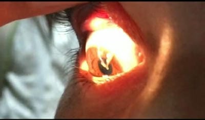
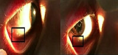
Ocular Thelaziasis is an arthropod-borne, zoonotic disease of the eye affecting the conjunctival sac, lacrimal duct, and lacrimal gland caused by a nematode of the genus Thelazia. Thelazia species are transmitted by different species of Muscidae, which are a family of flies with worldwide distribution. The present study reports a case of human ocular Thelaziasis in a 13-year-old female patient. Species Thelazia callipaeda was confirmed based on microbiological examination. The patient was treated with anti-helminthic drugs and was relieved of the symptoms without recurrence.

Keywords: Thelazia callipaeda, Thelaziasis, Zoonotic disease

Corresponding Author How to Cite this Article To Browse
Anchal Tripathi, Senior Resident, Department of Ophthalmology, LLRH, GSVM Medical College, Kanpur, Uttar Pradesh, India.
Email:
Tripathi A, Bhalla S. Wriggly creatures coming out of eye: case report on human ocular thelaziasis. Trop J Ophthalmol Otolaryngol. 2020;5(8):250-253.
Available From
https://opthalmology.medresearch.in/index.php/jooo/article/view/172

Manuscript Received Review Round 1 Review Round 2 Review Round 3 Accepted
2020-11-06 2020-11-16 2020-11-28 2020-12-18
Conflict of Interest Funding Ethical Approval Plagiarism X-checker Note
No Nil Yes 5%

© 2020 by Anchal Tripathi, Sonali Bhalla and Published by Siddharth Health Research and Social Welfare Society. This is an Open Access article licensed under a Creative Commons Attribution 4.0 International License https://creativecommons.org/licenses/by/4.0/ unported [CC BY 4.0].

Introduction

Thelaziasis is an ocular arthropod-borne disease of the eye infesting the conjunctival sac, lacrimal duct, and gland caused by a nematode of the genus Thelazia [1]. In general, patients with Thelaziasis are asymptomatic or present with excessive lacrimation. The two Thelazia species associated with Thelaziasis in humans are Thelazia callipaeda (Oriental eye worm) and Thelazia californiensis (California eye worm) [2]. The only confirmed vector for T callipaeda is the fruit fly, Phortica variegate (Diptera, Drosophilidae, Steganinae), which feeds on ocular secretions of their definitive hosts, including cats, dogs, horses, cattle, and humans [3].

The vectors feed on the conjunctival secretions of the infected hosts and ingest the first-stage larva along with it. The larva matures into their third larval (infective) stage in 2-3 weeks within the vector [4]. When the vector (fly) feeds in the eye of a new definitive host, including humans, the larvae enter the conjunctival sac of the host eye and mature into the adult stage, after two molts, within one month. Both adult and larval stages may live in conjunctival or lacrimal apparatus in the definitive host and cause conjunctivitis, keratitis, lacrimation, and ocular discharge. The adult females release first-stage larvae into the lacrimal secretions by ovoviviparity [5,6]. Thus, the life cycle continues.

Clinical manifestations of Thelaziasis range from ocular pruritis, lacrimation, epiphora, exudative conjunctivitis, or corneal edema to keratitis and corneal ulceration in severe cases leading to blindness [7]. Clinical diagnosis of Thelaziasis in animals and humans may be difficult and misleading. A confirmed diagnosis of Thelaziasis is usually made by the ophthalmologists based on visualization of the parasite, the eggs, or the larvae under a microscope.

Morphological differentiation between T callipaeda and T californiensis is based on the numbers of pre-and post-cloacal papillae in the male and the position of the vulva in the female [8]. In general, the male T callipaeda worm has 8-10 pairs of preanal papillae and 5 pairs of postanal papillae, whereas the male T californiensis worm has 6-7 pairs of preanal papillae and 3 pairs of postanal papillae [2]. Although many drugs have a promising effect, mechanical removal of the parasite is the only definitive treatment for ocular Thelaziasis [1].

Case Report

The present study reports a rare case of human Thelaziasis in a 13-year-old female child from Kanpur district, Uttar Pradesh, India. The patient complained of a foreign body sensation in her right eye. She also gave the history of recovering wriggly creatures from her right eye, with her bare fingers. The child is from a poor family and lives in an area where humans and animals live close to each other.

Her family owns a buffalo. On examination, it was noted that the child was repeatedly rubbing her right eye. The patient was noted to have excessive lacrimation and mucoid discharge (Figure 1). No purulent discharge or trauma was noted. Visual acuity was 20/20 in both eyes. Slit-lamp examination revealed small, whitish, motile worms wriggling over her bulbar conjunctiva (Figure 2).

A total of 10 worms were removed from the patient’s right eye, using sterile forceps (Figure 3). They were collected in a 0.9% NaCl bottle and were sent for microbiological examination. All 10 worms were identified as T callipaeda, 3 were male, and 7 were female. The worms were translucent, thin thread-like, and measured approximately 11-13mm in length in females and 8-10mm in length in males.

The female worm had a buccal capsule, mouth opening with a long muscular esophagus, and a conical tail with the vulval opening located at the anterior portion of the esophagointestinal junction. The male worm also possessed a similar buccal cavity but distinct esophageal-intestinal junction and characteristic curved tail end with shorter spicule with pre-and post-cloacal papillae. Based on these morphological features, the collected nematodes were identified as T callipaeda.

The patient was prescribed albendazole 400mg tablet stat for deworming. Moxifloxacin 0.5% ophthalmic drops were prescribed at a frequency of 4 times per day to prevent secondary bacterial infections. CMC 1% eye drops were prescribed at a frequency of 4 times per day for added lubrication.

The patient was first followed up after three days, and thereafter every two weeks. She was relieved of her symptoms on her first follow up. No worms were recovered on further follow-up. The patient was followed up for 3 months to look for recurrence.


opthal_172_01.jpg

Fig-1: Copious discharge as a presenting feature.

opthal_172_02.jpg

Fig-2: White slimy worms visible over the sclera.

opthal_172_03.jpg

Fig-3: Manual removal of thelazia worms through straight non-toothed forceps under a slit lamp.

Discussion

Ocular infections pose a critical threat internationally. Since the eyes are exposed directly to the environment, they are vulnerable to infectious diseases caused by vector-borne parasites. Timely identification and treatment are critical to prevent complications. Thelaziasis, like other ocular parasitic infections, occur more commonly in low socioeconomic regions and rural communities where humans live near animals [12]. Unhygienic environments and animal waste attract a variety of flies near the house, similar to our case. New-borns and young children, as noted in our case report, are at risk due to increased risk of exposure and because they are unable to protect themselves from the flies sitting on their eyes.

Although our case presented early, the late or neglected presentation may lead to severe diseases such as corneal ulceration and blindness [9]. The 2 species known to cause human Thelaziasis are T callipaeda and T californiensis. Thelazia callipaeda is commonly found in Asia, whereas T californiensis is commonly found in the United States [11].

In a recent study, a third species, Thelazia gulosa, has been reported to cause human thelaziasis in the United States [12]. Thelazia callipaeda was first documented by Railliet and Henry in 1910 [2]. The only confirmed vector for T callipaeda is P variegate (Diptera, Drosophilidae, Steganinae) [4]. However, Amiota okadai is also considered to be a vector of this parasite in China (summarised by Otranto et al [13].

Phortica flies are attracted by the eye secretions of humans, animals (such as dogs), and carnivores, and they feed on these secretions. Due to their zoophilic feeding habit, Phortica flies act as a potential vector of T callipaeda [13]. The presence of P variegate is known in countries such as Buzau, Giurgiu, Constana, Caras- Severin, Mehendinti, Timis, Maramaures, Ialomita, and Teleorman [14].

It has been reported that treatment for canine infection of T callipaeda with topical organophosphates, 1% moxidectin, or a formulation containing 10% imidacloprid and 2.5% moxidectin is effective [1]. However, the mechanical removal of parasites in humans remains the only curative option [1].

Nevertheless, due to localization of the nematode, Thelaziasis can be treated by direct application of drugs into the eyes. Patients with an intraocular infestation with T callipaeda have been successfully treated with a pars plana vitrectomy [10]. Michalski (1976) found that 2ml of levamisole injected into the subconjunctival sac was more effective than levamisole given orally [10].

In the case of T californiensis, removal of the worm will resolve the symptoms, and irrigation with Lugol’s iodine or 2%-3% boric acid is recommended immediately after worm removal or for the parasites that are in the lacrimal ducts where they cannot be removed manually [10].

Levamisole, either orally at 5 mg/kg or 2 ml injection into the conjunctival sac, has been recommended before the availability of ivermectin [12].


A dose of 2 mg/kg ivermectin given subcutaneously has also been shown to cure similar infestations in Asia and Europe [10,12]. Because our case is a young child and manual removal of worms with a cotton swab or forceps was sufficient [12], levamisole and ivermectin were not used. However, the patient was under close follow-up every two weeks.

Also, moxifloxacin ophthalmic drops were given to the patient to prevent a secondary bacterial infection because the initial few worms were removed by the patient herself using the bare finger. Currently, there is no vaccine for thelaziasis [10].

Thus, the prevention of human thelaziasis should include stringent control of the fly vector, including the use of bed nets to protect young children while they are sleeping and maintaining eye hygiene [1].

Conclusion

Thelaziasis is an emerging zoonotic disease in low socioeconomic regions. It occurs in rural communities where humans live near animals, similar to our case. Also, this condition is likely underreported, and a detailed epidemiological study on this parasite is needed to shed more light on the present status of this infection in India.

Public health programs that promote hygienic environments free from insect vectors, such as flies, are vital to prevent ocular infections. In infected patients, removal of the worm may provide definitive treatment and prevent complications.

Our case highlights the importance of awareness of this emerging infection, which is critical for timely diagnosis among ophthalmologists and clinicians to prevent further devastating ocular complications.

Reference

  1. Otranto D, Dutto M. Human thelaziasis, Europe. Emerg Infect Dis. 2008;14(4)647-649.
  2. Viriyavejakul P, Krudsood S, Monkhonmu S, Punsawad C, Riganti M, Radomyos P. Thelazia callipaeda- a human case report. Southeast Asian J Trop Med Public Health. 2012;43(4)851-856.
  1. Mihalca AD, D’Amico G, Scurtu I, Chirilă R, Matei IA, Ionică AM. Further spreading of canine oriental eyeworm in Europe- first report of Thelazia callipaeda in Romania. Parasite Vectors. 2015;8(1)48.
  2. Otranto D, Traversa D. Thelazia eyeworm- an original endo- and ecto-parasitic nematode. Trends Parasitol. 2005;21(1)1-4.
  3. De NV, Le TH, Chai JY. The first human case of Thelazia callipaeda infection in Vietnam. Korean J Parasitol. 2012;50(3)221-223.
  4. Otranto D, Lia RP, Buono V, Traversa D, Giangaspero A. Biology of Thelazia callipaeda (Spirurida, Thelaziidae) eyeworms in naturally infected definitive hosts. Parasitol. 2004;129(5)627.
  5. Vieira L, Rodrigues FT, Costa Á, Diz-Lopes D, Machado J, Coutinho T, et al. First report of canine ocular thelaziosis by Thelazia callipaeda in Portugal. Parasit Vectors. 2012;5(1)124.
  6. Naem S. Thelazia species and conjunctivitis, In- Pelikan, Z, editors, Conjuctivitis- A Complex and Multifaceted Disorder. Croatia, Europe- INTECH open science/open minds. 2011;201–32;ch-13.
  7. Yang YJ, Liag TH, Lin SH, Chen HC, Lai SC. Human thelaziasis occurrence in Taiwan. Clin Exp Optom. 2006;89(1)40-44.
  8. Bradbury RS, Breen KV, Bonura EM, Hoyt JW, Bishop HS. Case report- Conjunctival infestation with Thelazia gulosa- a novel agent of human thelaziasis in the United States. Am J Trop Med Hyg. 2018;98(4)1171–1174.