E-ISSN:2456-6454
P-ISSN:2581-4907
RNI:MPENG/2017/74152

Case Report

Atresia

Tropical Journal of Ophthalmology and Otolaryngology

2020 Volume 5 Number 6 July-August
Publisherwww.medresearch.in

A case report: bilateral choanal atresia in a nine-year-old female child

Shaik W.1*, Aradhyula D.2, Rao Yedluri S.3
DOI: https://doi.org/10.17511/jooo .2020.i06.09

1* Wahid Shaik, Senior Resident, Department Of Otorhinolaryngology, NRI Academy of Sciences, Guntur, Andhra Pradesh, India.

2 Dinesh Aradhyula, Post-Graduate, Department Of Otorhinolaryngology, NRI Academy of Sciences, Guntur, Andhra Pradesh, India.

3 Satya Prabhakar Rao Yedluri, Professor and Head of The Department, Department Of Otorhinolaryngology, NRI Academy of Sciences, Guntur, Andhra Pradesh, India.

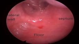
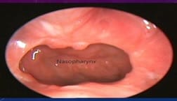
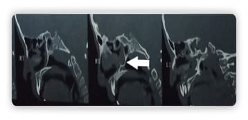
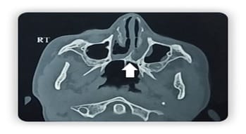
Choanal atresia is a rare congenital malformation of the nasal cavity characterized by obliteration of posterior choanae. It can be unilateral or bilateral. Bilateral choanal atresia is one of the life-threatening conditions and survival up to adulthood is rare A 9-year-old female presented to our department with complaints of a bilateral nasal block, nasal discharge, snoring, anosmia, and mouth breathing. Diagnostic nasal endoscopy and computerized tomography of the nose and paranasal sinuses revealed bilateral choanal atresia. Transnasal endoscopic choanoplasty was performed and discharged on postoperative day 7. Postoperative follow up on two weeks showed significant improvement in symptoms and endoscopy revealed bilateral patent posterior choanae. The child with bilateral choanal atresia surviving up to nine years of age is rare. In contrast to unilateral choanal atresia, bilateral choanal atresia is a diagnostic and therapeutic emergency. Diagnostic nasal endoscopy and computerized tomography help in planning surgery. Minimally invasive endoscopic choanoplasty has replaced the transpalatal approach.

Keywords: Choanal atresia, Congenital, Choanoplasty

Corresponding Author How to Cite this Article To Browse
Wahid Shaik, Senior Resident, Department Of Otorhinolaryngology, NRI Academy of Sciences, Guntur, Andhra Pradesh, India.
Email:
Shaik W, Aradhyula D, Yedluri S R P. A case report: bilateral choanal atresia in a nine-year-old female child. Trop J Ophthalmol Otolaryngol. 2020;5(6):168-171.
Available From
https://opthalmology.medresearch.in/index.php/jooo/article/view/150

Introduction

Choanal atresia (incidence 1 in 7000 live births), is a rare congenital malformation of the nasal cavity characterized by obliteration of posterior choanae. It can be unilateral or bilateral, bony, membranous, or mixed, complete or, incomplete. It is believed to be secondary to the persistence of the nasobuccal membrane. Only nine cases were

reported up to date [1,2].

Respiratory distress is a common presenting symptom. Bilateral choanal atresia will present as an acute respiratory emergency at birth as newborns are obligate nasal breathers. The inability to pass nasal catheters suggests the diagnosis, which can be confirmed with flexible nasal endoscopy, and computerized tomography of the nose and paranasal sinuses to determines the


Manuscript Received Review Round 1 Review Round 2 Review Round 3 Accepted
2020-06-27 2020-07-18 2020-07-31 2020-08-26
Conflict of Interest Funding Ethical Approval Plagiarism X-checker Note
No Nil Yes 7%

© 2020 by Wahid Shaik, Dinesh Aradhyula, Satya Prabhakar Rao Yedluri and Published by Siddharth Health Research and Social Welfare Society. This is an Open Access article licensed under a Creative Commons Attribution 4.0 International License https://creativecommons.org/licenses/by/4.0/ unported [CC BY 4.0].

extent and nature of the choanal atresia. The two most common techniques for choanal atresia repair are the transnasal and transpalatal approaches. Transpalatal technique is used in patients with significant craniofacial anomalies. Bilateral choanal atresia is rare compared to unilateral. The case is about a nine-year-old bilateral choanal atresia, describing the clinical features, how it was investigated, and managed.

This work is written by following the SCARE criteria [3].

Case Report

A nine-year-old female child presented to our department with complaints of bilateral nasal obstruction, nasal discharge, snoring, anosmia, mouth breathing, episodes of bluish discoloration of the body, frequent episodes of cold. No history of trauma or nasal surgery. History of cleft lip correction on the right side at the age of 3 years.

The child was born of consanguineous marriage. On Clinical examination, the findings are ectropion of the right lower lid, hypertelorism, hypoplasia of the right maxilla, high arched palate, crowding of teeth. The examination of the ear showed a normal external auditory canal with a normal tympanic membrane. Functional examination of the ear is normal

opthal_150_01.jpg

opthal_150_02.jpg

opthal_150_03.jpg

Fig-1: Preoperative and postoperative endoscopic images with 00 endoscopes.

The systemic examination is within normal limits. Diagnostic nasal endoscopy showed a deviation of the septum to the left with pooling of secretions in both nasal cavities. Mucosa covered defect between the septum and lateral wall (Figure 1). The scope could not be passed into the nasopharynx on both sides. Computerized tomogram of the nose and paranasal sinuses showed soft tissue densities in the posterior choana and bony thickening extending from the posteromedial wall of the maxilla to the vomer suggesting [mixed type] bilateral choanal atresia with adenoid hypertrophy (Figures 2 and 3).

opthal_150_04.jpg

Fig-2. CT Nose and PNS sagittal view showing thick bony atretic plate.

opthal_150_05.jpg

Fig-3: CT scan Nose and PNS Axial view showing thickening of the vomer extending to the posteromedial wall of maxilla.


Transnasal endoscopic choanoplasty is performed resecting the vomer, posterior end of the nasal septum with the help of Kerrison bone punch. Posterior choanal repermeabilization was achieved using microdebrider. Patency checked to bypass a rubber catheter into the nasopharynx. Deviation of the septum to the left is corrected along with the removal of adenoids using endoscopic transnasal microdebrider. Bilateral nasal packing is done. Nasal packs were removed on postoperative day four and, the patient was advised nasal douching with normal saline and discharged on a postoperative day 7. The patient came to review after two weeks to the outpatient department for follow up, showing significant symptomatic improvement and diagnostic nasal endoscopy revealed bilateral patent choana.

Discussion

Choanal atresia - a congenital malformation is characterized by the obliteration of posterior choana. It affects 1 in 5000-7000 births and is associated with other congenital abnormalities in 50% cases. Female predominance has been reported to choanal atresia with a male to female ratio being 1:2. 2 out of 3 cases are unilateral. The bilateral form represents a diagnostic and therapeutic neonatal emergency. It is associated with other congenital abnormalities in 50% of the cases [2,4].

Choanal atresia is a common component of congenital disorders such as CHARGE, Treacher Collins syndromes. CHARGE is an abbreviation for coloboma of the iris, heart defects, atresia of choana, growth retardation, genital abnormalities, ear abnormalities. The detailed cellular and molecular mechanism underpinning the etiology and pathogenesis remain elusive [5]. It is believed to be secondary to the persistence of the nasobuccal membrane.

The last study discovered that mutations in retinol dehydrogenase 10, which perturbs vitamin A metabolism and retinoid signaling, exhibit fully penetrant choanal atresia [5]. The revelation of congenital bilateral choanal atresia in adulthood remains exceptional in partial or neglected forms. In our case, it is reported that a congenital bilateral choanal atresia. The atretic plate thickness varies from 1 to 12mm [4].

The choanal atresia takes various aspects on the anatomical plane: Single or bilateral atresia; bone, membranous or mixed; Symmetrical Shrinkage or

not, predominant, or not on one wall or the other [6]. A mirror is placed near one nostril and then the other to observe for fogging on the mirror in the existence of patent choanae.

Usually, bilateral choanal atresia patients present with bilateral complete nasal obstruction, respiratory distress, cyclic respiratory obstruction. Transnasal endoscopic choanoplasty was performed in our patient. It began with choanal repermeabilization using a microdebrider followed by enlargement of the posterior part of the nasal cavity intersecting the vomer bone whose resection of its posterior border is the most efficient gesture to obtain a large and stable orifice. The reconstitution of the mucous plane is done to prevent restenosis.

Repair of choanal atresia has evolved over the past few decades. The open trans palatal technique has given way to minimally invasive endoscopic repair [7]. Bilateral nasal packing was done, and removal is done on a postoperative day four followed by nasal douching with normal saline. The initial review showed minimal crusting, and endoscopy revealed a patent posterior choana. Six months follow up showed well patent choana with significant improvement of symptoms. Of the nine documented cases of adult bilateral choanal atresia in literature, only one patient had restenosis, requiring revision surgery [8-10]. Aksoy et al. used mitomycin C after choanoplasty to prevent stenosis [9].

The role of a stent is controversial. Some investigators advocate its supremacy [8-12].

Conclusion

The bilateral choanal atresia is considered incompatible with life. Early surgical intervention is required in the neonatal period. Adult bilateral choanal atresia is one of the rare entities. Nasal endoscopy and computerized tomography preoperatively help to guide the surgical procedure. Transnasal endoscopic choanoplasty is the therapeutic choice. The purpose of this review is to provide a comprehensive clinical update on choanal atresia and to identify areas of future study.

Reference

  1. Chaudhary N, Jain A, Kapoor R, Motwani G. Bilateral complete choanal atresia in an adult woman--managed with nasal endoscopes. J Indian Med Assoc. 2010;108(2)109-110.

  1. Panda NK, Simhadri S, Ghosh S. Bilateral choanal atresia in an adult- is it compatible with life?. J Laryngol Otol. 2004;118(3)244-245.
  2. Agha RA, Fowler AJ, Saeta A, Barai I, Rajmohan S, Orgill DP, et al. The SCARE Statement- Consensus-based surgical case report guidelines. Int J Surg. 2016;34;180-186.
  3. Ramsden JD, Campisi P, Forte V. Choanal Atresia, and Choanal Stenosis. Otolaryngol Clin North Am. 2009;42(2)339-352.
  4. Kurosaka H, Wang Q, Sandell L, Yamashiro T, Trainor, PA. Rdh10 loss-of-function and perturbed retinoid signaling underlie the etiology of choanal atresia. Hum Mol Genet. 2017;26(7)1268–1279.
  5. Anderhuber W, Stammberger H. Endoscopic surgery of uni-and bilateral choanal atresia. Auris Nasus Larynx. 1997;24(1)13-19.
  6. Bangiyev JN, Govil N, Sheyn A, Haupert M, Thottam PJ. Novel application of steroid eluting stents in choanal atresia repair- a case series. Ann Otol Rhinol Laryngol. 2017;126(1)79-82.
  1. Tinoco P, Pereira JCO, Lourenço Filho RC, Brito TSC, Pereira BM, Carrara VL, et al. Bilateral choanal atresia in a 34-year-old patient. Int Arch Otorhinolaryngol. 2010;14(4)481-484.
  2. Aksoy F, Demirhan H, Yildirim YS, Ozturan O. Bilateral choanal atresia in an adult - management with mitomycin C and without stents- a case report. Cases J. 2009;2(1)9307.
  3. Verma RK, Lokesh P, Panda NK. Congenital bilateral adult choanal atresia undiagnosed until the second decade- How we did it. Allergy Rhinol (providence). 2016;7(2)82-84.
  4. Coniglio JU, Manzione JV, Hengerer AS. Anatomic findings and management of choanal atresia and the CHARGE association. Ann Otol Rhinol Laryngol. 1988;97(5 Pt 1)448-453.
  5. Gawai SA, Mail NJS. Bilateral Complete Congenital Choanal Atresia in an Adult Managed Endoscopically with Mucosal Flaps without Stenting. Otolaryngology Online J [Internet]. 2016;6(3).