E-ISSN:2456-6454
P-ISSN:2581-4907
RNI:MPENG/2017/74152

Research Article

Technique

Tropical Journal of Ophthalmology and Otolaryngology

2021 Volume 6 Number 3 May-June
Publisherwww.medresearch.in

Comparison of conjunctival free autograft and Rotational flap technique in primary pterygium surgery: Visual changes and safety profiles

Chaurasia P.1, Chaurasia V.2*
DOI: https://doi.org/10.17511/jooo.2021.i03.03

1 Pawan Chaurasia, Assistant Professor, Department of Opthalmology, Peoples College of medical sciences, Bhopal, Madhya Pradesh, India.

2* Vaishali Chaurasia, Associate Professor, Department of Obstetrics and Gynecology, Gandhi Medical College, Bhopal, Madhya Pradesh, India.

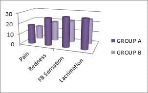
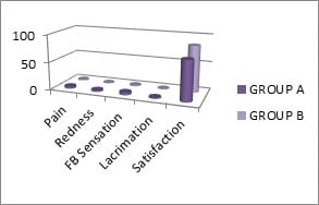
Aims and objectives: To evaluate the postoperative symptomatic comfort, visual changes, complication and rate of recurrence in free conjunctival autograft and rotational flap technique in primary pterygium surgery. Materials & Methods: This prospective study was conducted in 60 eyes of 60 patients, presented with primary progressive nasal pterygium larger than 1mm causing symptomatic discomfort, astigmatism and cosmetic disfigurement. Patients were divided into two groups randomly. In group A, patients with pterygium excision with free conjunctival autograft and group B, pterygium excision with rotation flap technique were included. Post-operative day 1 symptomatic comfort (symptoms and signs), graft stability, corneal clarity and any complication were noted. Visual acuity (VA), auto-refracto keratometer measurements and detailed biomicroscopic examinations, were performed preoperatively and postoperatively at 1month and 3 months. Results: Most of the patients in our study were in the middle age group of 40-49 years (41.6%). Out of 60 patients, 35 were males (58.4%) and 25 were females (41.6%). Patients with outdoor activities had a higher prevalence of pterygium (78.4%). The incidence of pterygium was more in the right eye (60%) than left eye (40%). Of the 60 patients, Grade I, II and III pterygium was 20%, 56.7% and 23.3% in group A and 16.7%, 63.3% and 20% in group B respectively. The mean symptomatic score was statistically significantly higher for group A for each factor (P<0.05). In the 3rd month, the overall patient’s satisfaction score was significantly higher in group B (P<0.05). Conclusion: Both surgical techniques were equally effective in terms of visual acuity, astigmatism and recurrence. The patient’s satisfaction score was significantly higher in the rotation flap technique group.

Keywords: Pterygium, Conjunctival free autograft, Rotational flap

Corresponding Author How to Cite this Article To Browse
Vaishali Chaurasia, Associate Professor, Department of Obstetrics and Gynecology, Gandhi Medical College, Bhopal, Madhya Pradesh, India.
Email:
Chaurasia P, Chaurasia V. Comparison of conjunctival free autograft and Rotational flap technique in primary pterygium surgery: Visual changes and safety profiles. Trop J Ophthalmol Otolaryngol. 2021;6(3):50-55.
Available From
https://opthalmology.medresearch.in/index.php/jooo/article/view/149

Manuscript Received Review Round 1 Review Round 2 Review Round 3 Accepted
2021-04-10 2021-04-20 2021-04-30 2021-05-04 2021-05-10
Conflict of Interest Funding Ethical Approval Plagiarism X-checker Note
No Nil Yes 5%

© 2021 by Pawan Chaurasia, Vaishali Chaurasia and Published by Siddharth Health Research and Social Welfare Society. This is an Open Access article licensed under a Creative Commons Attribution 4.0 International License https://creativecommons.org/licenses/by/4.0/ unported [CC BY 4.0].

Introduction

Pterygium is a triangular wing-shaped, fibro-vascular tissue derived from conjunctiva encroaching the cornea[1],which lead to cosmetic and visual deterioration secondary to induced astigmatism and visual axis involvement. Although several theories have been associated with etiology, its pathology remains controversial [2]. It is more common in the countries with relatively high exposure to ultraviolet radiation, the hot and dusty climates, and those living in rural areas and some occupational groups like agriculture workers, labourers, drivers, welders and carpenters. In recent studies, tropical residence and ultraviolet radiation exposure have been implicated as risk factors in the etiopathogenesis of pterygium formation. [3,4]. There are various surgical techniques for pterygium excision like simple excision, pterygium excision with conjunctival free autograft or rotational flap, having their own sets of advantages and disadvantages. Simple surgical excision (the bare sclera) is a simple and common procedure used to remove a pterygium. However, the bare sclera is associated with a rate of recurrence as high as 80%.(5).To reduce the recurrence, Mitomycin C (MMC), amniotic membrane and conjunctival transplantation are used after simple surgical excision. [6,7].

The use of Mitomycin C is associated with many side effects such as congestion, corneal epithelial defect, corneal perforation and sclera thinning. [8,9]. Conjunctival tissue is used either as a free autograft or rotation flap after simple pterygium excision to reduce surgical recurrence and to avoid the side effects of MMC.[10]. Of the two transplant procedures, the free conjunctival autograft procedure has lesser recurrence and complication rates but requires training by qualified surgeons and has a longer surgical time. [11]. In this procedure, the graft is obtained from supero-temporal/nasal bulbar conjunctiva, leaving behind a bare scleral area of higher dimension which heals in a longer time, which leads to post-operative irritation and discomfort to the patient. Conjunctival rotation flap surgery, on the other hand, is a relatively simple procedure to master and surgical time is also short.[12]. The objectives of this study were to compare symptomatic comfort, visual outcome, recurrence and complications in free conjunctival autograft and rotation flap techniques following primary pterygium surgery.

Materials & Methods

This was a prospective interventional study conducted in the department of ophthalmology, Chirayu Medical College & Hospital Bhopal for a period of 2 years from April 2017 to March 2019 after clearance from the institutional ethical committee. 60 eyes of 60 primary pterygium patients attending outpatient department and indoor in the department were included in the study. Informed and written consent was taken from all the patients regarding the procedure, outcome and follow up.

Inclusion criteria: Patients above 18 years of age, with a progressive nasal pterygium with corneal encroachment, which is causing discomfort and irritation, chronic inflammation, visual disability or cosmetic disfigurement.

Exclusion criteria

  • History of prior pterygium surgery
  • Pterygium with concurrent ocular surface and lid disease
  • Pseudo-pterygium or collagen vascular disease
  • Cases with a history of glaucoma or glaucoma suspect
  • Patients not giving consent

A detailed history was taken and demographic data were recorded. The corneal encroachment of the pterygium was measured using a slit-lamp biomicroscope by a single observer. If a pterygium was ≤2 mm inside the cornea, it was classified as Grade 1, 2-4 mm as Grade 2, and ≥4 mm as Grade 3. The patients were divided into two groups namely A and B randomly by a computer-generated number. In group A (n-30) patients underwent pterygium excision followed by free conjunctival autograft and in group B (n-30) pterygium excision followed by rotation flap technique was performed. All procedures were performed by a single surgeon experienced in pterygium surgery and conjunctival transplant procedures.

After instilling topical anaesthesia eye was cleaned with betadine solution followed by sterile draping, the eye was exposed using a universal eye speculum. Subconjunctival anesthesia (epinephrine and lidocaine) was administered under the pterygium body and on the superior part of the conjunctiva with a 26 and half-gauge needle.


The pterygium was cut near the limbus by Wescott’s scissors, the head of the pterygium was dissected from the surface of the cornea and subconjunctival fibrous tissue was removed completely. Any residual fibrous tissue was removed with the use of a 15 number blade. Cautery was used minimally in case of abnormal bleeders in the bare scleral area.

For the free conjunctival auto-grafting procedure, the graft tissue was harvested from supero-temporal/nasal bulbar conjunctiva, approximately 1 mm larger than the area of the bare sclera. The flap was dissected, completely freed from the underlying Tenon's tissue and transposed over the bare area. For conjunctival rotational flap procedure, the flap was created from the superior bulbar conjunctiva, rotated to the bare area aligning the limbal to limbal end of the flap with the scleral bed. In both the procedures uniform pressure was applied on the graft using 2 iris repositors, so that the graft gets adhered to the underlying sclera with the help of the blood coagulum.

After the application of a 0.5% Moxifloxacin with 0.1% Dexamethasone eye ointment, the eye was closed with a sterile pad. Surgical time was noted in both procedures. Postoperatively antibiotic steroid combination (Moxiloxacin+Loteprednol) eye drops in tapering doses (starting with four times a day for 4 weeks) and tears substitutes were prescribed to all patients (4 times/day) for 4 weeks on discharge. Postoperative follow up was done on the first postoperative day then at one week, one month and three months after surgery for symptomatic comfort, visual outcome, recurrence and complications.

Symptomatic comfort, graft stability, corneal clarity and any complication were noted postoperative day 1 and at day 7, whereas visual acuity, auto-refracto keratometer measurements and biomicroscopic examinations were performed at 1 month and 3 months follow up. At each follow-up visit, a questionnaire was filled by the patients, for grading pain, redness, foreign body (F.B) sensation and lacrimation into four grades.

The questionnaire was scored from (0 to 3) 0 - nothing; 1 -mild; 2 - moderate; 3 -severe. Additionally, in the 3rd month, the overall satisfaction with the procedure was recorded as four grades 0 -unsatisfied; 1 - low satisfaction, 2 -moderate satisfaction and 3 - highly satisfied. Both the groups were compared for ocular signs and symptoms and overall satisfaction.

Recurrence was defined as fibrovascular proliferation invading the cornea more than 1.5 mm.

Statistical analysis: Statistical analysis was performed using Statistical Package for Social Sciences (SPSS) version 20. Data were presented as mean, standard deviation and range. Snellen vision values were converted into log MAR values for statistical analysis. P-values less than 0.05 were considered statistically significant.

Results

A total of 60 patients were selected randomly and divided into two groups A (30) and B (30). Most of the patients in our study were in the middle age group of 40-49 years (41.6%). Out of 60 patients, 35(58.4%) were males and 25(41.6%) were females Patients with outdoor activities had a higher prevalence of pterygium (78.4%). The incidence of pterygium was more in the right eye (60%). Grade I, II and III pterygium 20%, 56.7% and 23.3% in group A and 16.7%, 63.3% and 20% in group B respectively. (Table-1)

Table-1: Patient demographics.

  GROUP A (N-30) GROUP B (N-30)
Age of presentation (in years)
<20-29
30-39
40-49
50-59
> 60
4
9
12
3
2
6
8
13
2
1
Gender
Males
Females
18
12
17
13
Occupation
Outdoor
Indoor
23
7
24
6
EYE
Right eye
Left eye
17
13
19
11
Grade of pterygium
Grade-1
Grade-2
Grade-3
6
17
7
5
19
6

Table-2: Presents the surgical time, complications and recurrence. The mean surgery time in the conjunctival free flap was 20-25 minutes and that in rotational flap techniques was15-20 minutes. Graft edema occurred in 16.7% of cases in group A and 10% cases in group B. Graft edema resolved gradually within 15days of surgery with steroids eye drops.


The displaced graft was reported on postoperative day 1 in two patients of group A and one patient of group B. reposition was done under topical anaesthesia with the help of iris repositors. persistent epithelial defect occurred in one patient with a rotational flap which was managed conservatively. Conjunctival granuloma formation was observed in one patient with free autograft, and was treated conservatively. The recurrence rate was 6.7% (n=2) in group A and 3.4% (n=1) in group B at the 3 months follow up. There were no major complications like graft necrosis, corneal thinning, scleral thinning or necrosis.

Table-2: Comparison of Surgical time, Complications and Recurrence.

  GROUP A GROUP B
Surgical time (in minutes) 20-25minutes 15-20minutes
Postoperative complications
Type of complication
1.Graft edema
2.Displaced graft
3.Persistent epithelial defect
4.Granuloma formation
5
2
-
1
3
1
1
-
Recurrence 2 1

Table 3: Presents the pre and post-operative visual acuity and astigmatism. The pre and postoperative mean BCVA (Log MAR) were compared at 1 month and 3 months. In group A preoperative mean BCVA of 0.48 ± 0.42 was significantly improved to 0.36 ± 0.44 (P < 0.001) at 1 month and 0.36 ± 0.44 (P < 0.001) at 3months after surgery. In group B preoperative mean BCVA of 0.46 ± 0.39 was significantly improved to 0.37 ± 0.41 (P < 0.001) and 0.34 ± 0.41 (P < 0.001) at 1 month and 3months postoperative, respectively.

The preoperative mean astigmatism (Refractive cylinder) was 1.38 ± 1.56 D in group A and 1.41 ± 1.63D in group B. In group A, postoperative mean astigmatism reduced to 0.87 ±0.92 D and 0.84±0.75D at 1month and 3 months respectively. In group B postoperative mean astigmatism reduced to 0.92 ±0.86 D at 1month and 0.87±0.79D at 3 months.

The changes in corneal astigmatism were statistically significant at 1 month and 3rd month. The preoperative mean corneal astigmatism was 2.33 ± 1.62 D and 2.41 ± 1.71D in group A and group B respectively. In group A postoperative mean corneal astigmatism reduced to 1.38±0.92 D at 1month and 1.12±0.81 D at 3rd-month post-surgery.

In group B postoperative mean astigmatism reduced to 1.42 ±0.96 D and 1.23±0.94 D at 1month and 3rd month respectively.

Table 3: Comparison of Pre and Postoperative Visual Acuity and Astigmatism

  GROUP A GROUP B
Preoperative Visual acuity (logMAR) 0.48±0.42 0.46±0.39
Astigmatısm(Refractive cylinder) 1.38 ±1.56D 1.41 ±1.63D
Corneal Astigmatism 2.33 ±1.62D 2.41 ±1.71D
Postoperative At 1 month Visual acuity (logMAR) 0.36±0.44 0.37±0.41
Astigmatısm(Refractive cylinder) 0.87 ±0.92D 0.92 ±0.86D
Corneal Astigmatism 1.38 ±0.92D 1.42 ±0.96D
Postoperative At 3rd month Visual acuity (logMAR) 0.36±0.44 0.34±0.41
Astigmatısm(Refractive cylinder) 0.84±0.75D 0.87±0.79D
Corneal astigmatism 1.12 ±0.81D 1.23 ±0.94D

Figure 1 & 2: Show the post-operative means score of symptoms and signs on day 1, at1 month and at the end of 3rd month. The mean symptomatic score was statically significant higher for group A for each factor (P<0.05). In the 3rd month, the overall patient’s satisfaction score was significantly higher in group B (P<0.05).

opthal_149_01.jpg

Figure 1: Postoperative symptoms and signs at day 1 in both groups.

opthal_149_02.jpg


Figure 2: Postoperative symptoms and signs at 1month in both groups.

There was a statistically significant difference in the scores of the postoperative pain, redness, foreign body sensation and lacrimation (p<0.05) in the two groups.

Discussion

Usually the pterygium is an asymptomatic wing-shaped fleshy mass, but it can present as symptomatic discomfort including redness, inflammation, foreign body sensation and in progressive cases, it can deteriorate vision as it encroaches the cornea with obscuring the visual axis of the cornea and inducing astigmatism. The Indications for pterygium surgery include patient discomfort and irritation, chronic inflammation, visual disability or cosmetic disfigurement.

Primary pterygium surgery aims to improve symptomatology, preserve or improve visual acuity and maintain ocular surface regularity and prevent a recurrence. The pterygium surgery is a minor surgical procedure, but if not done properly, it may lead to recurrence. Cases who underwent rotational flap surgery had better symptomatic comforts and overall satisfaction than Group A (p<0.05) whereas graft stability was comparable in both the groups. In both groups autologous blood coagulum was used for conjunctival grafting in pterygium surgeries. It has fewer postoperative discomforts, easy to use and avoid suture related complications.

In our study we found that visual acuity improved after pterygium excision surgery followed by conjunctival grafting in both groups. BCVA significantly improved from 0.48 ± 0.42 and 0.46±0.39 preoperatively to 0.36 ± 0.44 and 0.34 ± 0.41 postoperatively in group A and B respectively (at 3 months; P <0.0001). Maheshwari S [13]. concluded that visual acuity significantly improves in all grades of pterygium after pterygium excision surgery (P <0.05). The reason for the improvement in visual acuity after pterygium surgery was lesser induced astigmatism and clearance of visual axis, previously obscured by pterygium.[8].

Several previous researchers have found that pterygium excision surgery significantly reduces pterygium induced corneal astigmatism. Mohite et al [14]. found a significant reduction in mean corneal astigmatism after pterygium surgery from 3.046 ± 1.20 D to 1.486 ± 0.63D (P<0.001).

Similar results were obtained in our study where a significant reduction in mean corneal astigmatism after pterygium surgery was reported. Maheshwari S [13]. and Avisar R et al 15 found that successful pterygium surgery reduced the pterygium induced refractive astigmatism and improved the visual acuity. Lindsay and Sullivan [16]. also found a significant correlation between pterygium surgery and improvement in visual acuity.

Accurate size of graft and transplant over the bare scleral area leads to lower recurrence of pterygium. We used graft tissue that was at least 1 mm larger as compared to the bare scleral site in all cases. In our study the recurrence rate was 6.7% (2 patients) in group A and 3.4% (1patient) in group B at the end of 3rd month.

In the study by Aslan [12]. rate of recurrence was comparable following auto conjunctival graft and rotational flap surgery (P=0.46). Ti et al [17]. also suggested that the size of the graft might be important in preventing the recurrence of pterygium.

Conclusion

Both surgical techniques were equally effective in terms of visual acuity, astigmatism and recurrence; however patient satisfaction score was significantly higher in patients undergoing surgery with the rotational flap method.

Reference

01. Di Girolamo N, Chui J, Coroneo MT, Wakefield D. Pathogenesis of pterygia- role of cytokines, growth factors, and matrix metalloproteinases. Prog Retin Eye Res. 2004 Mar;23(2)195-228. doi: 10.1016/j.preteyeres.2004.02.002 [Crossref][PubMed][Google Scholar]

02. Coroneo MT, Di Girolamo N, Wakefield D. The pathogenesis of pterygia. Curr Opin Ophthalmol. 1999 Aug;10(4)282-8. doi: 10.1097/00055735-199908000-00011 [Crossref][PubMed][Google Scholar]

03. Di Girolamo N, Coroneo MT, Wakefield D. UVB-elicited induction of MMP-1 expression in human ocular surface epithelial cells is mediated through the ERK1/2 MAPK-dependent pathway. Invest Ophthalmol Vis Sci. 2003 Nov;44(11)4705-14. doi: 10.1167/iovs.03-0356 [Crossref][PubMed][Google Scholar]


04. Golu A, Gheorghişor I, Bălăşoiu AT, Baltă F, Osiac E, Mogoantă L, Bold A. The effect of ultraviolet radiation on the cornea - experimental study. Rom J Morphol Embryol. 2013;54(4)1115-20. [Crossref][PubMed][Google Scholar]

05. Riordan-Eva P, Kielhorn I, Ficker LA, Steele AD, Kirkness CM. Conjunctival autografting in the surgical management of pterygium. Eye (Lond). 1993;7( Pt 5)634-8. doi: 10.1038/eye.1993.146 [Crossref][PubMed][Google Scholar]

06. Alpay A, Uğurbaş SH, Erdoğan B. Comparing techniques for pterygium surgery. Clin Ophthalmol. 2009;3;69-74. [Crossref][PubMed][Google Scholar]

07. Narsani AK, Nagdev PR, Memon MN. Outcome of recurrent pterygium with intraoperative 0,02% mitomycin C and free flap limbal conjunctival autograft. J Coll Physicians Surg Pak. 2013 Mar;23(3)199-202. [Crossref][PubMed][Google Scholar]

08. Rubinfeld RS, Pfister RR, Stein RM, Foster CS, Martin NF, Stoleru S, et al. Serious complications of topical mitomycin-C after pterygium surgery. Ophthalmology. 1992 Nov;99(11)1647-54. doi: 10.1016/s0161-6420(92)31749-x [Crossref][PubMed][Google Scholar]

09. Safianik B, Ben-Zion I, Garzozi HJ. Serious corneoscleral complications after pterygium excision with mitomycin C. Br J Ophthalmol. 2002 Mar;86(3)357-8. doi: 10.1136/bjo.86.3.357 [Crossref][PubMed][Google Scholar]

10. Mahdy MA, Bhatia J. Treatment of primary pterygium- Role of limbal stem cells and conjunctival autograft transplantation. Oman J Ophthalmol. 2009 Jan;2(1)23-6. doi: 10.4103/0974-620X.48418 [Crossref][PubMed][Google Scholar]

11. Koranyi G, Seregard S, Kopp ED. The cut-and-paste method for primary pterygium surgery- long-term follow-up. Acta Ophthalmol Scand. 2005 Jun;83(3)298-301. doi: 10.1111/j.1600-0420.2005.00465.x [Crossref][PubMed][Google Scholar]

12. Aslan L, Aslankurt M, Aksoy A, Ozdemir M, Yüksel E. Comparison of wide conjunctival flap and conjunctival autografting techniques in pterygium surgery. J Ophthalmol. 2013;209401. doi: 10.1155/2013/209401 [Crossref][PubMed][Google Scholar]

13. Maheshwari S. Effect of pterygium excision on pterygium induced astigmatism. Indian J Ophthalmol. 2003 Jun;51(2)187-8. [Crossref][PubMed][Google Scholar]

14. Mohite U S, N B Dole, S S Jadhav. "Effectiveness of pterygium surgery on corneal astigmatism". Med Pulse Int J Ophthalmol. 3(2017)12-17. [Crossref][PubMed][Google Scholar]

15. Avisar R, Mekler S, Savir H. [Effect of pterygium excision on keratometric readings]. Harefuah. 1994 Jan 16;126(2)63-5,112,111. [Crossref][PubMed][Google Scholar]

16. Lindsay RG, Sullivan L. Pterygium-induced corneal astigmatism. Clin Exp Optom. 2001 Jul;84(4)200-203. doi: 10.1111/j.1444-0938.2001.tb05026.x [Crossref][PubMed][Google Scholar]

17. Ti SE, Chee SP, Dear KB, Tan DT. Analysis of variation in success rates in conjunctival autografting for primary and recurrent pterygium. Br J Ophthalmol. 2000 Apr;84(4)385-9. doi: 10.1136/bjo.84.4.385 [Crossref][PubMed][Google Scholar]