E-ISSN:2456-6454
P-ISSN:2581-4907
RNI:MPENG/2017/74152

Research Article

Demographic

Tropical Journal of Ophthalmology and Otolaryngology

2020 Volume 5 Number 3 March
Publisherwww.medresearch.in

Demographic and clinical profile of venomous snake bite-related ocular manifestations among the paediatric age group in a tertiary care hospital

Jana S.1, Ray S.2*, Banerjee M.3
DOI: https://doi.org/10.17511/jooo.2020.i03.04

1 Subhasis Jana, Senior Resident, Department of Ophthalmology, Burdwan Medical College and Hospital, Burdwan, West Bengal, India.

2* Soumya Ray, M.B.B.S., M.S.(PGT), Junior Resident, Department of Ophthalmology, Burdwan Medical College and Hospital, Burdwan, West Bengal, India.

3 Mousumi Banerjee, Head of the Department, Department of Ophthalmology, Burdwan Medical College and Hospital, Burdwan, West Bengal, India.

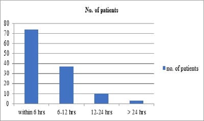
Background: The children having high case fatality and working children (10-14 years) actually belong to the high-risk group of snakebite cases. There is a significant lack of knowledge of the clinical-epidemic profile of snakebite cases in the pediatric age group. Objectives: To determine the ocular manifestations as well as the magnitude of long-standing visual impairment following snake bite among the pediatric population. Methods: A prospective observational cross-sectional study was conducted on all the venomous snake bite patients admitted in the pediatric ward of our institution. Bed side ophthalmological evaluation initially by recording visual acuity, ocular position, extraocular movements and anterior segment evaluation were done. Response to treatment and the residual ocular morbidity were also recorded. The collected data were analysed using appropriate statistical methods. Results: A total of 124 patients were presented to the pediatric department and included in the study. It was observed that males (75.81%) are a more common victim of snakebite whereas lower limb (89.50%) being the common site of snake bite. Total no of patients: 15 (12.09%) patients had ocular manifestations, dimness of vision is the most common symptom of neurotoxic snake bite followed by ptosis, external ophthalmoplegia, diplopia, optic neuritis, and significant persisting visual impairment. Conclusion: Early treatment and close monitoring will help to reduce ocular morbidity. Health education to increase awareness about the hazards of snakebite, early hospital referral, and effective treatment will reduce the global burden of human suffering inflicted by this neglected tropical disease.

Keywords: Venomous snake bite, Ocular manifestations, Ocular morbidity

Corresponding Author How to Cite this Article To Browse
Soumya Ray, M.B.B.S., M.S.(PGT), Junior Resident, Department of Ophthalmology, Burdwan Medical College and Hospital, Burdwan, West Bengal, India.
Email:
Jana S, Ray S, Banerjee M. Demographic and clinical profile of venomous snake bite-related ocular manifestations among the paediatric age group in a tertiary care hospital. Trop J Ophthalmol Otolaryngol. 2020;5(3):78-83.
Available From
https://opthalmology.medresearch.in/index.php/jooo/article/view/136

Manuscript Received Review Round 1 Review Round 2 Review Round 3 Accepted
2020-03-08 2020-03-18 2020-03-23 2020-02-28
Conflict of Interest Funding Ethical Approval Plagiarism X-checker Note
No Nil Yes 9%

© 2020 by Subhasis Jana, Soumya Ray, Mousumi Banerjee and Published by Siddharth Health Research and Social Welfare Society. This is an Open Access article licensed under a Creative Commons Attribution 4.0 International License https://creativecommons.org/licenses/by/4.0/ unported [CC BY 4.0].

Introduction

Observing the importance of snakebite in human health, WHO has included snake bite in the list of “Neglected Tropical Diseases” [1]. According to recent WHO statistics in India alone, as many as 2.8 million people are bitten by snakes, and 46,900 people die from snakebite every year [2]. Due to the practices of traditional remedies in rural India data seems to be underestimated [3]. Having high rate of prevalence, Rural West Bengal in India showed an average annual mortality rate of 16 per 100,000 population [4]. The venomous snakes most often encountered in India are the Indian spectacled cobra (Najanaja), common krait (B. caeruleus), Russell's viper (Daboia russelli) and the Saw scaled viper (Echiscarinatus). Though ocular complications following snake bite are usually rare, still some of them are reported in the literature as ophthalmoplegia, uveitis, glaucoma, central retinal artery occlusion, macular infarction, optic neuritis, vitreous hemorrhage, penetrating eye injury, globe necrosis, cortical infarction and endogenous endophthalmitis [5-11]. Most of the literature state about the ocular complications of snakebite envenomation among adults. But the under 5 children having high case fatality and working children (10-14 years) actually belong to the high-risk group of snakebite cases [2]. There is a significant lack of knowledge of the clinical-epidemic profile of snakebite cases in the pediatric age group. With this background, this hospital-based cross-sectional study among pediatric patients was undertaken to evaluate the demographic and clinical profile of venomous snake bite-related ocular manifestations.

Materials and Methods

This was a prospective observational cross-sectional study conducted on all the venomous snake bite patients admitted in the pediatric ward of Burdwan Medical College and Hospital, a tertiary care center with rural set up in the last 1 year (May 2018 to April 2019). After taking approval of the Institutional Ethics Committee, the study was initiated. All snakebite patients admitted in the Dept. of Paediatrics, of this hospital, were examined and only cases of envenomation with ocular manifestations were included in the present study. The venomous bite was defined by the presence of signs and symptoms of local and/or systemic toxicity.

Local toxicity was defined by the presence of a local reaction in the form of swelling, bleeding from fang marks, cellulitis, or necrosis. Clinical features such as bleeding from mucocutaneous sites, systemic bleeding, intravascular hemolysis including disseminated intravascular coagulation (DIC), or a deranged laboratory coagulation profile which includes 20 minutes whole blood coagulation time, bleeding time, clotting time, APTT (Activated partial thromboplastin time) anytime during hospital stay indicated a vasculogenic bite. The neurotoxic bite was considered in case of paresthesias, taste and smell abnormalities, ptosis, cranial nerve palsy, general flaccidity, or even respiratory paralysis. Polyvalent anti-snake venom (ASV) and other supportive therapy were instituted as per hospital protocol.

Bedside ophthalmological evaluation was done initially by recording visual acuity, ocular position, extraocular movements, and anterior segment evaluation were done. The fundus examination was done after dilating with tropicamide eye drops 0.8% when possible.

Later when the patient became ambulatory detailed evaluation was done in the ophthalmology outpatient department. Response to treatment and the residual ocular morbidity was also recorded.

A detailed proforma was prepared for the purpose of recording the following data as observations from the present study. Socio-demographic profiles of the snakebite victims, the pattern of snakebites, time and place of occurrence of the snakebite, trends of snakebite, site, and human activity at the time of the bite, details of the circumstances leading to the event; and post-bite factors including the outcomes and other relevant epidemiological data, etc. The ocular morbidity profile, as well as laboratory profile, included blood profile and other important information was obtained from the records. The collected data were analyzed using appropriate statistical methods.

Results

A total of 124 patients were presented to the pediatric department and included in the study. As Table 1 shows the demographic profile of study participants, it can be observed that males (75.81 %) were a more common victim of snakebite whereas lower limb (89.50 %) being the common site of snake bite.


Table-1: Demographic profile of patients of this study: n=124.

Sex Male 94 (75.81%)
Female 30 (24.19 %)
Religion Hindu 45 (36.29 %)
Muslim 64 (51.61 %)
Others 15 (12.1 %)
Education/literacy Literate 119 (95.97 %)
Illiterate 05 (04.03 %)
Site of bite Lower limb 111 (89.50 %)
Upper limb 13 ( 10.50 %)

Mean age of study patients: 7.52±2.83years.

74 patients presented within 6 hours, 37 presented within 6-12 hours, 10 presented within 12-24 hours and 3 presented > 24 hours. Neurotoxic bites: 40 patients (32.25 %), Vasculotoxic bites: 84 patients (67.74 %).

opthal_136_01.jpg

Fig-1: Mean time between snakebite and AVS loading.

Ocular manifestations: Out of 124 pediatric snake bite cases only 15 (12.09 %) patients had ocular manifestations. As from Table 2, it can be observed dimness of vision being the most common symptom of neurotoxic snake bite followed by ptosis, external ophthalmoplegia, diplopia, optic neuritis, and significant persisting visual impairment.

Table-2: Ocular manifestations of neurotoxic snake bite 4 patients (26.67% of all patients that had ocular manifestations. N= 4) (here data are not mutually exclusive).

Ocular manifestations No. of patients
Dimness of vision 4
Ptosis 4
External ophthalmoplegia 3
Diplopia 2
Optic neuritis 1
Significant persisting visual impairment 1

From Table 3 it can be observed that the ocular manifestations of vasculotoxic snake bite. Sub conjunctival hemorrhage(90.90%) being the commonest followed by retinal hemorrhage, vitreous hemorrhage, lid edema, hyphema, anterior uveitis, and significant persisting visual impairment.

Table-3: Ocular manifestations of vasculotoxic snake bite, 11 patients (73.33 % of all patients that had ocular manifestations, N= 11) (here data are not mutually exclusive).

Ocular manifestations No. of patients
Subconjunctival hemorrhage 10 (90.90 %)
Retinal hemorrhage 6
Vitreous hemorrhage 3
Lid edema 5
Hyphema 2
Anterior uveitis 2
Significant persisting visual impairment 2

Discussion

The present study highlights the burden of ocular manifestations of snakebite and its long-standing visual impairment in the pediatric population. Most of the literature state about the ocular complications of snakebite envenomation among adults. There is a significant lack of knowledge of the clinical-epidemic profile of snakebite cases in the pediatric age group. Out of 124 cases of snakebite that were admitted in the hospital during the study period, 15 cases (12.09 %) had ocular manifestations and were analyzed.

Snake venom is a complex mixture of several enzymes and proteins, toxic polypeptides, and inorganic components. it contains numerous toxins, and their combined action has a more potent effect than that of their individual effects. In general, venoms are described as either neurotoxic or hematotoxicity [12]. Cobra and krait are neurotoxic, whereas Viperidae species show characteristic vasculotoxic and haematotoxic features. Patil VC et al reported that 85.43% cases in their series were vasculotoxic and 14.56% were neurotoxic [13]. Males were more commonly affected than females. Brunda G et al reported 76% cases of snakebite to be males [2]. It was also observed that a male preponderance and higher incidence of vasculotoxic bites in our series.

Ocular manifestations were more common in vasculotoxic snake bite in the present study. The commonest finding is subconjunctival hemorrhage


( 90.90 %) which contradicts the findings of Das et al [14] were retinal and vitreous hemorrhage were noted to be the commonest. Vasculotoxic snake venom has several biologically active procoagulant enzymes that lead to rapid activation of factors V, X amongst other steps in the clotting cascade. This leads to the formation of fibrin cross-links, which are promptly lysed by the fibrinolytic system [15]. Eventually, this leads to disseminated Intravascular coagulation and coagulopathy that can cause sub-conjunctival and intraocular hemorrhage. Interestingly 2 cases of anterior uveitis following snakebite have been reported which also corroborates the findings of Das et al [14]. One case was acute, noted at presentation and the other case manifested 7 days post-admission to hospital. The cause of uveitis is unclear. This can be explained by the observations of different authors like Buttes GP et al and Kumar PK et al [16,17] reporting anterior uveitis may be due to the direct toxic effect of the snake venom. Here late-onset uveitis following a venomous bite is probably due to serum sickness-like response to horse ASV serum which is also explained by S Nayak et al [18].

In the present study, common ocular manifestations of neurotoxic snake bites were ptosis followed by external ophthalmoplegia, diplopia, and optic neuritis, in order of decreasing incidence. Seneviratne U et al [19] in their study of 56 patients with neurological manifestations of snakebite, reported that ptosis and ophthalmoplegia were the commonest neurological manifestations. Singh J et al [20] in an analysis of 33 venomous snake bites in a military operational area of north India reported 21 patients to be neurotoxic in nature. Ptosis, diplopia and blurred vision were common finding Neuromuscular paralysis occurs as a result of the blockade of neuromuscular transmission.

The neurotoxins have phospholipase A2 activity and hydrolyze phosphoglycerides thereby producing neuromuscular blockade by inhibiting the release of acetylcholine from the presynaptic membrane [21,22]. The extraocular muscles are especially susceptible to neuromuscular blockage because the ratio of nerve fibers to extra-ocular muscle fibers (1:6 to 1:12) is high compared to that seen in the large proximal limb muscles (1:2000) [23]. Therefore, a small amount of neurotoxin is capable of producing an increased effect on the extraocular and levator muscles. This explains the present study finding of ptosis and ophthalmoplegia to be the commonest features of neurotoxic envenomation.

Out of 15 cases that had ocular manifestations in the present study only 3 cases suffered significant visual impairment at the end of six months follow up. One patient had a persistent uniocular vitreous hemorrhage, the second patient had bilateral late-onset severe uveitis along with vitreous hemorrhage, and the third patient suffered from optic neuritis sequelae along with hemorrhage on the macula. The first patient refused any intervention in his eye.

Limitations of the study

As no study is without limitations so this also true for the present study. In developing countries like ours, snakebite victims may preferentially attend traditional healers and may not seek treatment at hospital thus the true burden of disease is not reflected here. Besides, the study duration is short, and the follow up was only for six months.

Conclusion

This study highlights the spectrum of ocular manifestations in venomous snake bites in a pediatric population. There is an urgent need to develop health intervention strategies specially for children to achieve more rapid access to antivenom in health facilities.

What does the study add to the existing knowledge?

Early treatment and close monitoring will help to reduce ocular morbidity. Health education to increase awareness about the hazards of snakebite, early hospital referral, and effective treatment will reduce the global burden of human suffering inflicted by this neglected tropical disease.

Author’s contribution

Dr. Subhasis Jana: Concept, study design

Dr. Soumya Ray: Manuscript preparation

Prof. (Dr.) Mousumi Banerjee: Manuscript preparation

Acknowledgment

The authors acknowledge the support of the clinicians and other staff of the Paediatrics department. The assistance from every member of the Ophthalmology department is highly appreciated.


Reference

01. Warrell DA, Gutiérrez JM, Calvete JJ, Williams D. New approaches and technologies of venomics to meet the challenge of human envenoming by snakebites in India. Indian J Med Res. 2013;138 (1)38-59. [Crossref][PubMed][Google Scholar]

02. Prevalence of snakebite envenoming. Available from [Article] [Crossref][PubMed][Google Scholar]

03. Mohapatra B, Warrell DA, Suraweera W, Bhatia P, Dhingra N, Jotkar RM, et al. Million Death Study Collaborators, Snakebite mortality in India- A nationally representative mortality survey. PLoS Negl Trop Dis. 2011;5(4)e1018. doi: 10.1371/journal.pntd.0001018 [Crossref][PubMed][Google Scholar]

04. Hati AK, Mandal M, De MK, Mukherjee H, Hati RN. Epidemiology of snake bite in the district of Burdwan, West Bengal. J Indian Med Assoc. 1992;90(6)145-147. [Crossref][PubMed][Google Scholar]

05. Bhalla A, Jain AP, Banait S, Jajoo UN, Kalantri SP. Central retinal artery occlusion- an unusual complication of snakebite. J venom Anim Toxins incl Trop Dis. 2004;10(3)311-314. doi: [Article][Crossref][PubMed][Google Scholar]

06. Takeshita T, Yamada K, Hanada M, Oda-Ueda N. Extraocular muscle paresis caused by snakebite. Kobe J Med Sci. 2003;49(1)11-15. [Crossref][PubMed][Google Scholar]

07. Menon V, Tandon R, Sharma T, Gupta A. Optic neuritis following snake bite. Indian J Ophthalmol. 1997;45(4)236-237. Available from: [Article][Crossref][PubMed][Google Scholar]

08. Singh J, Singh P, Singh R, Vig VK. Macular infarction following viperine snake bite. Arch Ophthalmol. 2007;125(10)1430-1431. doi: 10.1001/archopht.125.10.1430 [Crossref][PubMed][Google Scholar]

09. Hayreh SS. Transient central retinal artery occlusion following viperine snake bite. Arch Ophthalmol. 2008;126(6)870-871. doi: 10.1001/archopht.126.6.870-a [Crossref][PubMed][Google Scholar]

10. Rao BM. A case of bilateral vitreous haemorrhage following snake bite. Indian J Ophthalmol. 1977;25(2):1-2. Available from: [Article][Crossref][PubMed][Google Scholar]

11. Iqbal M, Khan BS, Ahmad I. Endogenous Endophthalmitis Associated with Snake Bite. Pak J Ophthalmol. 2009;25(2)114-116. [Crossref][PubMed][Google Scholar]

12. Law AD, Agrawal AK, Bhalla A. Indian common krait envenomation presenting as coma and hypertension- A case report and literature review. Emerg Trauma Shock. 2014;7(2)126-128. doi: 10.4103/0974-2700.130887 [Crossref][PubMed][Google Scholar]

13. Patil VC, Patil HV, Patil A, Agrawal V. Clinical Profile and outcome of envenomous snake-bite at tertiary care centre in western Maharashtra. Int J Med Public Health. 2011;4(1)28-38. doi: 10.5530/ijmedph.4.2011.7 [Crossref][PubMed][Google Scholar]

14. Das S, Samadder S, Jana S. Ocular Manifestation and Long-Standing Visual Impairment Following Venomous Snake Bite. J Evolution Med Dent Sci. 2015;4(46)7919-7923. doi: 10.14260/jem ds/2015/1153 [Crossref][PubMed][Google Scholar]

15. Tungpakorn N. Unusual visual loss after snakebite. J Venom Toxins Incl Trop Dis. 2010;16(3)519-523. doi: 10.1590/S1678-91992010 000300020 [Crossref][PubMed][Google Scholar]

16. Buttes GP, Ayan N, Cami G. Uveitis after snakebite. Arch Pediatr. 1996;3(8)832-833. doi: 10.1016/0929-693x(96)82175-0 [Crossref][PubMed][Google Scholar]

17. Kumar PK, Ahuja S, Kumar PS. Bilateral acute anterior uveitis and optic disc edema following a snake bite. Korean J Ophthalmol. 2014;28(2)186-188. doi: 10.3341/kjo.2014.28.2.186 [Crossref][PubMed][Google Scholar]

18. Nayak SG, Satish R, Nityanandam S, Thomas RK. Uveitis following anti-snake venom therapy. J Venom Toxins Trop Dis. 2007;13(1)13. doi: 10.1590/S1678-91992007000100010 [Crossref][PubMed][Google Scholar]

19. Seneviratne U, Dissanayake S. Neurological manifestations of snake bite in Sri Lanka. Journal of Postgrad Med. 2002;48(4)275-278. Available from: [Article][Crossref][PubMed][Google Scholar]

20. Singh J, Bhoi S, Gupta V, Goel A. Clinical profile of venomous snake bites in north Indian Military Hospital. J Emerg Trauma Shock. 2008;1(2)78-80. doi: 10.4103/0974-2700.43184 [Crossref][PubMed][Google Scholar]


21. Bawaskar HS, Bawaskar PH. Envenoming by the common krait (Bungarusbcaeruleus) and Asian cobra (Najanaja)- Clinical manifestations and their management in a rural setting. Wilderness Environ Med. 2004;15(4)257-266. doi: 10.1580/1080-6032(2004)015[0257:ebtckb]2.0.co;2 [Crossref][PubMed][Google Scholar]

22. Lewis RL, Gutmann L. Snake venoms and the neuromuscular junction. Semin Neurol. 2004;24(2)175-179. doi: 10.1055/s-2004-830904 [Crossref][PubMed][Google Scholar]

23. Kim HD, Jung MS, Kim SY. Exotropia caused by pit viper snakebite. J AAPOS. 2009;13(4)424-425. doi: 10.1016/j.jaapos.2009.04.018 [Crossref][PubMed][Google Scholar]