E-ISSN:2456-6454
P-ISSN:2581-4907
RNI:MPENG/2017/74152

Case Report

Vascularisation

Tropical Journal of Ophthalmology and Otolaryngology

2020 Volume 5 Number 2 February
Publisherwww.medresearch.in

A case report of oculofacial rosacea complicating to corneal infiltrates and vascularisation

Chanekar S.1*, P. S. Usgaonkar U.2, O Akarkar S.3
DOI: https://doi.org/10.17511/jooo.2020.i02.04

1* Samruddhi Chanekar, Junior Resident, Department of Ophthalmology, Goa Medical College, Bambolim, Goa, India.

2 Ugam P. S. Usgaonkar, Professor and Head, Department of Ophthalmology, Goa Medical College, Bambolim, Goa, India.

3 Shekhar O Akarkar, Senior Resident, Department of Ophthalmology, Goa Medical College, Bambolim, Goa, India.

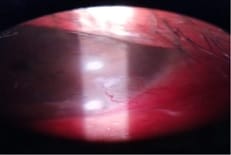
Purpose: To report a case of ocular rosacea with corneal infiltrates and vascularisation. Here we report a case of a 34-year-old male presenting with ocular rosacea with corneal infiltrates and vascularisation in the left eye with posterior blepharitis of both eyes. The clinical diagnosis was based on the facial findings of erythematous papulomacular lesions and telangiectasia. The patient was treated with systemic doxycycline and for ocular lesions treated with antibiotic steroids and lubricants. Ocular rosacea, if not treated on time, can worsen to the stage of corneal infiltrates and vascularisation.

Keywords: Ocular rosacea, Corneal infiltrates, Blepharitis

Corresponding Author How to Cite this Article To Browse
Samruddhi Chanekar, Junior Resident, Department of Ophthalmology, Goa Medical College, Bambolim, Goa, India.
Email:
Chanekar S, Usgaonkar UPS, Akarkar SO. A case report of oculofacial rosacea complicating to corneal infiltrates and vascularisation. Trop J Ophthalmol Otolaryngol. 2020;5(2):60-62.
Available From
https://opthalmology.medresearch.in/index.php/jooo/article/view/133

Manuscript Received Review Round 1 Review Round 2 Review Round 3 Accepted
2020-02-05 2020-02-16 2020-02-23 2020-02-28
Conflict of Interest Funding Ethical Approval Plagiarism X-checker Note
No Nil Yes 8%

© 2020 by Samruddhi Chanekar, Ugam P. S. Usgaonkar, Shekhar O Akarkar and Published by Siddharth Health Research and Social Welfare Society. This is an Open Access article licensed under a Creative Commons Attribution 4.0 International License https://creativecommons.org/licenses/by/4.0/ unported [CC BY 4.0].

Introduction

Rosacea is a chronic inflammatory disorder mainly involving midline facial skin and neck, with onset in the age group of 30-50 years, with preponderance in females than males. Skin involvement varies from flushing, erythema, telangiectasia, papules, and pustules to phymatous changes. Common ocular manifestations include meibomian gland dysfunction, lid telangiectasias, conjunctival hyperemia, blepharoconjunctivitis. Left untreated can lead to peripheral corneal vascularization, infiltrates, thinning and perforation [1]. Although ocular involvement is common, complications occur only in 6-18% of patients [2]. Ocular Rosacea is a relatively common condition that is often underdiagnosed and untreated. This case report is presented to stress on the importance of early diagnosis and treatment especially when cornea gets involved as it may lead to blindness if not treated in time.

Case Report

Thirty-Four years old male presented to the outpatient department with redness, watering, photophobia left eye for 2 weeks. There was a history of skin lesions over the face which had worsened for the last 3 weeks. Slit Lamp Examination of the left eye showed circumcorneal congestion, two subepithelial infiltrates, both measuring 2×2 mm, one perilimbal at around 5 o’clock and other paracentral at 4 o’clock with superficial corneal vascularization. (Figure 1) Both eyes had posterior blepharitis with eyelid margins showing telangiectasias and erythema. Both eyes had interpalpebral conjunctival hyperemia. Visual Acuity in the right eye was 6/9 and 6/24 in the left eye. The posterior segment of both eyes was normal. Schirmer’s test was 10 mm right eye and 7 mm in the left eye.

opthal_133_01.jpg

Fig-1: Left eye with corneal subepithelial infiltrates and vascularization.

opthal_133_02.jpg

Fig-2: Facial rosacea lesions.

There were papulomacular erythematous lesions involving the malar region, forehead, and nose. (Figure 2) The patient was diagnosed to have severe oculofacial rosacea. The patient was treated with systemic doxycycline 100 mg BD. Topically patient was treated with steroid eye drops (prednisolone acetate ophthalmic solution 1% ) 6 times a day that was tapered weekly, antibiotic eye drops (moxifloxacin 0.5% ) QID for two weeks, homatropine eye drops 2% TDS. Both eyes were put on lubricant eye drops 6 times. Warm compresses and the digital message was advised for both eyes.

After a two-week patient’s condition improved, with a decrease in conjunctival hyperemia and healing of corneal lesions. The patient was continued on the same medications.

Discussion

Rosacea is a chronic inflammatory skin disorder with yet unclear pathogenesis. The diagnosis of rosacea is usually clinical. Flushing, dentofacial erythema, and papules are most commonly observed. In 2002, the National Rosacea Society Expert Committee developed a classification system for rosacea, describing 4 different subtypes: erythematotelangiectatic, papulopustular, phymatous, and ocular rosacea [3].


Ocular and facial manifestations most commonly develop simultaneously, but at times ocular signs precede skin changes wherein it is frequently misdiagnosed. Both eyes are usually affected simultaneously, but unilateral or sequential involvements can occur. A common ocular manifestation of rosacea ranges from blepharitis, conjunctival injection, eyelid margin telangiectasias, meibomian gland dysfunction such as chalazion or stye to sight-threatening complications like corneal ulceration, vascularization, and perforation [4].

Corneal alterations are detected in 25–50% of patients with ocular rosacea and may range from mild punctate epithelial keratitis accompanying the blepharoconjunctivitis to corneal vascularization, infiltration, ulceration, and perforation. Peripheral corneal infiltrates/vascularization and superficial punctate keratitis, usually involving the inferior half of the cornea, are the most common corneal findings.

Characteristically, there is the vascular invasion of the peripheral cornea with subepithelial infiltrates along the advancing vascular border. These infiltrates can progress circumferentially or toward the central cornea. The infiltrates may be followed by corneal vascularization and may compromise the visual axis. This condition may also be associated with significant peripheral thinning and asymmetric astigmatism which also impairs vision. Severe corneal inflammation can lead to corneal melting and even perforation.

Duke-Elder reported that blepharitis in every case of rosacea eventually appears and sometimes spread to conjunctiva assuming diffuse hyperemic type and rarely nodular conjunctivitis. Nodular episcleritis near limbus and rosacea keratitis with marginal ulcer occurs as an extension of Rosacea. Conjunctivitis frequently accentuated in lower quadrants. This may be associated with punctate keratitis and later on subepithelial infiltrates develop. Numerous scars, horseshoe-shaped, or tongue-shaped, develop with the apex directed toward the center of the cornea [5].

The detailed etiology and pathophysiology of ocular and cutaneous rosacea remain unclear, although it is commonly thought of as an inflammatory process. Another hypothesis states that rosacea is dermal dystrophy where there occur degenerative changes in perivascular collagen causing small vessel dilatations and in due course incompetence of vessels.

Subsequent leakage of potentially inflammatory substances in perivascular space results in lymphoedema and formation of papules, pustules, and nodules. Studies have reported increased levels of interleukins 1a and b, matrix metalloproteinase 9 and collagenase 2 in tears of patients with ocular rosacea [1].

Oral doxycycline has been the mainstay of treatment for cutaneous and ocular rosacea. It has been shown to improve ocular disease and increase the tear break-up time [6]. It was found to decrease the concentration of MMP-8 and MMP-9 in the tear film [7,8]. The mainstay of treatment for blepharitis is lid hygiene [9]. Topical antibiotics have been used to alter the flora of the ocular surface in addition to topical steroids [10,11].

Rosacea is a prevalent disorder that may be disfiguring and cause significant social impairment, as well as various degrees of ocular morbidity, if not diagnosed and managed appropriately. Ocular manifestations may precede, follow or occur simultaneously with skin changes, but most commonly they develop jointly. The most If not treated it can lead to corneal complications like corneal infiltrates, vascularisation and perforation.

Reference

01. Audrey SC, Kathryn AC. Lid Inflammations, In- Albert DM, Jakobiec FA, Miller WJ (Eds), Principles and Practice of Ophthalmology- 3rd ed. Philadelphia- Elsevier. 2008;630-634. [Crossref][PubMed][Google Scholar]

02. Bowling B. Kanski’s Clinical Ophthalmology- A Systematic Approach. 8th ed, Edinburgh- Butterworth-Heinemann/Elsevier. 2007. [Crossref][PubMed][Google Scholar]

03. Wilkin J, Dahl M, Detmar M, Drake L, Feinstein A, Odom R, et al. Standard classification of rosacea-Report of the National Rosacea Society expert committee on the classification and staging of rosacea. J Am Acad Dermatol. 2002;46(4)584–587. doi: 10.1067/mjd.2002.120625 [Crossref][PubMed][Google Scholar]

04. Akpek EK, Merchant A, Pinar V, Foster CS. Ocular rosacea- Patient characteristics and follow-up. Ophthalmol. 1997;104(11)1863-1867. doi: 10.1016/S0161-6420(97)30015-3 [Crossref][PubMed][Google Scholar]


05. Duke Elder S. Diseases of outer eye, In- System of Ophthalmology. St Louis- CV Mosbey. Vol 8 Part-I, 1965;534-45. [Crossref][PubMed][Google Scholar]

06. Quarterman MJ, Johnson DW, Abele DC, Lesher JL Jr, Hull DS, and Davis LS. Ocular rosacea- Signs, symptoms, and tear studies before and after treatment with doxycycline. Arch Dermatol. 1997;133(1)49-54. doi: 10.1001/archderm.133.1.49 [Crossref][PubMed][Google Scholar]

07. Määttä M, Kari O, Tervahartiala T, Peltonen S, Kari M, Saari M, et al. Tear fluid levels of MMP-8 are elevated in ocular rosacea-treatment effect of oral doxycycline. Graefe's Arch Clin Exp Ophthalmol. 2006;244(8)957-962. doi: 10.1007/s00417-005-0212-3 [Crossref][PubMed][Google Scholar]

08. Sobrin L, Liu Z, Monroy DC, Solomon A, Selzer MG, Lokeshwar BL, et al. Regulation of MMP-9 activity in human tear fluid and corneal epithelial culture supernatant. Invest Ophthalmol Vis Sci. 2000;41(7)1703-1709. [Crossref][PubMed][Google Scholar]

09. Key JE. A comparative study of eyelid cleaning regimens in chronic blepharitis. CLAO J. 1996;22(3)209-212. [Crossref][PubMed][Google Scholar]

10. Odom R, Dahl M, Dover J, Draelos Z, Drake L, Macsai M, et al. Standard Management Options for Rosacea, Part 2- Options According to Subtype. Cutis- cutaneous medicine for the practitioner. 2009;84(2)97-104. [Crossref][PubMed][Google Scholar]

11. AlarfajK, Alzamil W. Spontaneous corneal perforation in ocular rosacea. Middle East Afr J Ophthalmol. 2010;17(2)186-188. doi: 10.4103/0974-9233.63070 [Crossref][PubMed][Google Scholar]