E-ISSN:2456-6454
P-ISSN:2581-4907
RNI:MPENG/2017/74152

Research Article

Abscess

Tropical Journal of Ophthalmology and Otolaryngology

2020 Volume 5 Number 1 January
Publisherwww.medresearch.in

Otogenic abscess: rarity and reality

Vishwanath Kale M.1*, C. Chhabria S.2, Sharma M.3, Subhash Gaikwad N.4
DOI: https://doi.org/10.17511/jooo.2020.i01.05

1* Meena Vishwanath Kale, Senior Resident, Department of ENT, T.N.M.C. & B.Y.L.Ch. Nair Hospital, Mumbai, Maharashtra, India.

2 Sanjay C. Chhabria, Assistant Professor, Department of ENT, T.N.M.C. & B.Y.L.Ch. Nair Hospital, Mumbai, Maharashtra, India.

3 Manisha Sharma, Junior Resident, Department of ENT, T.N.M.C. & B.Y.L.Ch. Nair Hospital, Mumbai, Maharashtra, India.

4 Ninad Subhash Gaikwad, Professor & HOD, Department of ENT, T.N.M.C. & B.Y.L.Ch. Nair Hospital, Mumbai, Maharashtra, India.

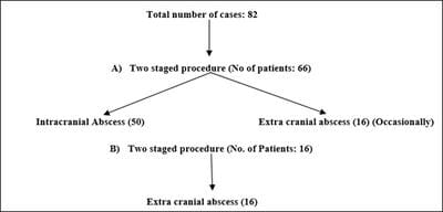
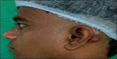
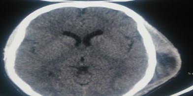
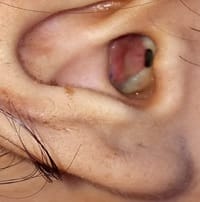
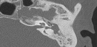
Introduction: Otogenic abscess is a complication commonly arising from active squamous chronic otitis media. It is rare (1:10000) but in fact are a dangerous reality to be managed promptly. Materials and Methods: A retrospective study of 82 cases of otogenic abscesses were studied over a period 24 years by simple random sampling. Cases of ear malignancy, previously operated mastoidectomy were excluded from study. All postop cases were analysed. Result: Most commonly affected were male (63.4%) in the second decade (34.1%) of life. Commonest bacterial infection was staphylococcus aureus (31.7%). Conclusion: Otogenic abscess though rare, are a reality and should be suspected when the patient with chronic ear discharge has high grade fever, severe headache and does not respond to best medical line of treatment. All these patients were subjected to imaging neurosurgical I&D of brain abscess followed by canal wall down tympano-mastoidectomy.

Keywords: Canal wall down tympano-mastoidectomy, Extracranial, Incision & Drainage (I &D), Intracranial, Otogenic abscess, Squamous active chronic otitis media

Corresponding Author How to Cite this Article To Browse
Meena Vishwanath Kale, Senior Resident, Department of ENT, T.N.M.C. & B.Y.L.Ch. Nair Hospital, Mumbai, Maharashtra, India.
Email:
Kale MV, Chhabria SC, Sharma M, Gaikwad NS. Otogenic abscess: rarity and reality. Trop J Ophthalmol Otolaryngol. 2020;5(1):22-29.
Available From
https://opthalmology.medresearch.in/index.php/jooo/article/view/127

Manuscript Received Review Round 1 Review Round 2 Review Round 3 Accepted
2020-01-10 2020-01-20 2020-01-25 2020-02-29
Conflict of Interest Funding Ethical Approval Plagiarism X-checker Note
No Nil Yes 7%

© 2020 by Meena Vishwanath Kale, Sanjay C. Chhabria, Manisha Sharma, Ninad Subhash Gaikwad and Published by Siddharth Health Research and Social Welfare Society. This is an Open Access article licensed under a Creative Commons Attribution 4.0 International License https://creativecommons.org/licenses/by/4.0/ unported [CC BY 4.0].

Introduction

Active squamous chronic otitis media (COM) is a synonym of cholesteatoma which leads to severe destruction with irreversible sequelae of intra or extracranial complications of otogenic origin [1]. Hence, an otogenic abscess is a rare clinical entity (1:10000) but required surgical emergency to be drained out to avoid complications [2].

As per our experience incidences of active squamous COM in spite of improved health education, socioeconomic status, resistance of organism to antibiotics and uncontrolled bacterial infection were noticed [1,3,4]. Due to in judicial use of latest antibiotics the disease has not yet completely regressed. Lifestyle changes attributed to metabolic diseases like diabetes mellitus and increased frequency of ear infections because of recurrent episodes of rhinosinusitis due to fitness activities like swimming, diving. Frequent use of ear plugs, air travels and recently increased use of cotton buds to clean the ear have also at times caused retraction of tympanic membrane.

Infection can spread from middle ear through preformed anatomical or pathological pathways like fracture of temporal bone, dehiscence of the floor of middle ear, non-union petro-squamous suture. Also spreads by 1) pathological implantation or invasion or metaplasia theory leads into bony erosion and osteitis of mastoid bone (tegmen tympani or Trautmann’s triangle) [5,6] or through 2) demineralization of bone or 3) venous thrombophlebitis i.e. connection between tributaries of dural veins with tributaries of Haversian canals; leads into meningitis, lateral sinus thrombophlebitis and finally leads into neurological instability of patients and 4) along with peri-arteriolar Virchow-Robin spaces into cerebral white matter. So squamous active COM can lead to lethal complications hence it’s a potentially serious disease [7].

Middle ear with mastoid erosion leads to collection of pus between the dura and cranium. It gets collected around the lateral sigmoid sinus and due to low host immunity leads to sigmoid sinus thrombosis which may extend down to involve internal jugular vein; it may further lead to complication of pulmonary embolism, ultimately leading to pulmonary infarct which due to early treatment is rare nowadays [5,8,9,10].

Less likely petrous apex abscess from mastoid bone erosion can lead to a trio of symptoms like I) Retro-orbital pain, II) lateral rectus palsy, III) Otorrhea which are collectively called Gradenigo’s syndrome [11].

First line treatment of otogenic abscess once diagnosed is medical to prevent impending complications. Treatment starts with hospitalization of patient, intravenous hydration therapy and broad-spectrum antibiotics. Clinically stabilization of neurological condition of patients with intracranial abscess may require lowering of raised cerebro-spinal fluid pressure with use of injection mannitol and extensive vital monitoring. Surgically incision and drainage of otogenic abscess is done at the earliest and once patient is stabilized, canal wall down tympano-mastoidectomy is treatment of choice [5].

Materials and Methods

Place of the study: Our tertiary care hospitals 1) K.E.M. Hospital, Parel and 2) B.Y.L. Ch Nair Hospital, Mumbai Central, also 3) Dr R. N. Cooper Hospital over a period of 27 years from 1993 to 2019.

Type of study: Retrospective analytical study

Sampling method: Simple random sampling

Sample collection: 82 cases of age group of 10 to 60 years

Inclusion criteria: cases who presented with squamous active COM with otogenic abscess

Exclusion criteria: Patients with mastoid malignancy, fibrous dysplasia, congenital atresia of EAC.

Statistical methods: Microsoft office 2007 was used to make tables. Descriptive statistics like mean and percentages were used to interpret the data and conclude the results.

Results

In this study, our main aim was to give a safe and dry ear to all patients who had squamous active COM diagnosed on clinical ENT evaluation with High Resolution Computed Tomography of Temporal bone (HRCT), CT with MRI brain and wherever necessary CSF Study and fundoscopy was done to rule out papillary edema [12,13]. Patient had undergone medical along with surgical management.


Following surgery options: as per patient’s medical treatment and clinical stability:

opthal_127_01.jpg

Fig-1: Surgery options

One staged procedure: Patients who were stable, underwent incision and drainage of extracranial abscess along with canal wall down tympano-mastoidectomy at the same sitting. In each case; pus for culture and sensitivity test (SCAST) collected and post-operatively patients were treated as per SCAST report.

Two staged procedure: Those who had intracranial abscess and unstable were treated by the two stage procedure i.e. initially intracranial abscess drainage with cyst wall excision by neurosurgeon through craniotomy or burr-hole surgery to avoid intracranial spread from infected origin i.e. mastoid followed by canal wall down tympano-mastoidectomy [14].

Sex distribution: Out of 82 patients 52 were male (63.4%) and 30 were female (36.5%). Males were more commonly affected than females [5].

Age distribution: In the present study, any age can be affected but most commonly the age of disease presentation was second decade (34.1%) followed by first decades (26.8%) [15]. While in age group of 31-40 years, 41-50 years and 51-60 years were also affected with number of patients were 12(14.6%), 11(13.4%) and 09 (10.9%) respectively.

Clinical Presentation:

Table-1: Clinical presentation.

Clinical features Extrcranial Intracranial
Otorrhoea 32 50
Otalgia 32 36
Deafness 28 44
Fever 26 42
Headache 10 44
Tinnitus 18 16
Signs of meningeal irritation 0 39
Vomiting 0 30
Post-aural swelling (Fig-2) 25 06
Papilloedema 0 15
Nystagmus 0 14
Altered sensorium 0 7
Vertigo 2 5
Facial palsy 6 11
Neck swelling 5 0
Sagging of external auditory canal 6 4
Gradenigo's syndrome 0 3

opthal_127_02.jpg

Fig-2: Left post-aural swelling suggestive of left mastoid abscess.

Most commonly observed cases were presented with foul smelling otorrhea with hearing loss (mixed or sensory-neural). Patients presented with signs of intracranial extension were about 50 (60.9%) cases and those with extra-cranial abscess were 32 (39.0%) cases.

Bacteriology: Bacterial infections are usually primary focus of infections which is detectable in pre-operative otorrhea and intraoperative granulation tissue histopathology. Medical treatment started as per SCAST report.

Table-2: Bacteria commonly involve in etiology

Bacteriology No. of patients Percentage
Staphylococcus aureus 26 31.7
Pseudomonas aeruginosa 21 25.6
Klebsiella aerogenes 14 17.0
Mycobacteria tuberculosis 09 10.9
Proteus mirabilis 06 7.31
Streptococcus viridans 04 4.87
Escherichia coli 02 2.43

Staphylococcus aureus (31.7%) and pseudomonas aeruginosa (25.6%) are mainly involve in pathology of disease [16]. In developing country, mycobacterium tuberculosis (10.9%) should keep in mind as infective pathology (Table 2).

Following surgery proposed: A] Two Staged Procedure: First incision and drainage of abscess followed by a week later canal wall down tympano-


mastoidectomy.

1) Patients with intracranial abscess :50

Table-3: Intracranial abscess: Two staged management

Abscesses No of cases Percentages (%)
Cerebral 18 21.9
Cerebellar 12 14.6
Meningitis 07 8.53
Extra-dural 06 7.31
Sigmoid sinus thrombosis 05 6.09
Petrous apex abscess 02 2.43

For meningitis, sigmoid sinus thrombosis and petrous apex abscess; medical management is the first line treatment. If medical line of treatment of sigmoid sinus thrombosis fails then surgical exploration with removal of thrombus and packing of cavity with gelfoam to block the vessel [17].

It was found (Table 3) that most commonly cerebral abscess (21.9%) in temporo-parietal region and then cerebellar abscess (14.6%). In these cases patients underwent abscess drainage with cyst wall excision through craniotomy or burr-hole surgery. After one week of stability and antibiotic coverage, patients had undergone canal wall down tympano-mastoidectomy.

2) Patients with extracranial abscess: 16

Table-4: Extracranial abscess: Two staged management

Abscesses No of cases Percentages (%)
Subperiosteal 08 9.75
Bezold’s 05 6.09
Zygomatic 02 2.43
Citelli’s 01 1.21

While in extracranial abscess, subperiosteal abscess were 9.75% and less number of Citelli’s abscess only 1.21% (Table 4). Extracranial abscess breakthrough mastoid bone and spread into subcutaneous plane (Figure 3) lead into postaural tender swelling.

opthal_127_03.jpg

Fig-3: Computed Tomography of Brain suggestive of complications left mastoid abscess in subcutaneous plane.

b] One staged Procedure: Incision and drainage along with canal wall down tympano-mastoidectomy at the same time.

Extra-cranial abscess as Luc’s abscess (fig:3) were 16(19.5%) cases were surgically managed with abscess drainage along with canal wall down tympano-mastoidectomy in a single stage surgery.

opthal_127_04.jpg

Fig-4: Luc’s Abscess

Abscess of posterior wall of external ossicious meatus through mastoid antrum

Table-5: Intra-operative Findings

Findings No of cases Percentages
Ossicular chain destruction 76 92.6
Posterior wall EAC erosion 74 90.2
Dural plate erosion 43 52.4
Dehiscence of facial canal 25 30.4
Sinus plate erosion 10 12.1
Sigmoid sinus thrombosis 05 6.09
Labyrinthine fistula of LSC (Figure 5) 04 4.87

opthal_127_05.jpg


Fig-5: HRCT Temporal bone: labyrinthine fistula: lateral semicircular canal dehiscence with soft tissue embedded in middle ear and mastoid cavity.

Table 6: Post-operative complications.

Complications No of cases Percentages
Discharging cavity only 10 12.1
Facial nerve palsy 04 4.88
Post-aural fistula 02 2.43

In the present study, there was no mortality but had raised morbidity due to intracranial abscess. In 12(14.63%) cases, patients required revision surgery for management of complications. Facial nerve palsy could not be corrected as they were irreversibly damaged by the disease.

Discussion

In the study of 82 cases of otogenic abscess who presented in our department, the discussion is based on clinical presentations, intra-operative findings and their management.

Squamous active COM with complications is more in developing countries [18]

In the study majority cases were between 10-30 years of age with maximum incidence in second decade (34.1%) followed by first decade (26.8%). Youngest being 12 years and oldest being 58 years. Males were predominantly affected (63.4%) with common presentation of foul smelling otorrhea occasionally blood tinged with hearing loss.

The bacterial etiology was mostly staphylococcus aureus (31.7%), pseudomonas aeroginosa (25.6%) and mycobacterium tuberculosis (10.9%) is also a common entity.

In the present study, the incidence of intracranial complications (60.9%) is more as compared to extracranial (39%) complications. Most common intracranial abscess was cerebral (21.9%) abscess while subperiosteal abscess (9.07%) was common in extracranial abscess.

All the 50 patients with intracranial abscesses had fundoscopy examination and graded accordingly - Grade I papilloedema was commonest 35 cases (42.6%), Grade II papilloedema was seen in 9 cases (10.9%) and Grade III papilloedema was seen in only 1 case (1.21%) of cerebral abscess. No patient showed Grade IV and V papilloedema [14].

After imaging (HRCT Temporal bone and MRI brain is the investigation of choice) and audiometry (was not possible in all patients due to their unstable condition) planned for surgery. Appropriate consent for unplanned facial nerve palsy, non-improvement of hearing and need for post-operative cleaning of mastoid cavity is explained.

The management depends on whether the abscess is intracranial or extracranial. In the present study, two staged procedure was preferred for both intracranial as well extracranial abscess as described above. Intracranial abscess drainage with cyst wall excision by craniotomy or burr hole surgery was preferred as it is not possible to excise the abscess wall through tympanomastoid approach which may lead to recurrence and also to avoid otogenic spread of infection into intracranial space.

Most commonly in ossicular chain destruction (92.6%); incus (lenticular and long process), followed by stapes suprastructure erosion. In certain cases ossicles were reshaped and implanted to improve degree of hearing. Post-operatively patient advised with hearing aid in case of decreased hearing.

Defects in the posterior wall of external auditory canal (EAC) in 90.2% of cases were converted to canal wall down procedure.

The current study have found dural plate, sinus plate defect and labyrinthine fistulae (4.87%) due to disease. Labyrinthine fistulae of lateral semicircular canal were only present and repaired intraoperatively by using bone dust and closing with temporalis fascia graft.

6.09% cases of sigmoid sinus thrombosis initially treated with conservative management (antibiotics) and if no response then drained out with wide bore needle or with surgical blade [19]. Role of anticoagulant is unclear [20].

Labyrinthine fistula (Figure 5) is the erosion of endochondral layer without perilymph leak is also seen as a complication of squamous COM [21]. It may due to choleasteatoma or inflammatory mediator of choleastetoma matrix [22]. It is usually done in two stage; initially possible cholesteatoma is removed and matrix at fistula site allow to change into cholesteatoma pearl. After three months, cholesteatoma pearl removed from the fistula and is sealed with bone dust and covered temporalis fascia graft [23,24]. At times the patient may be required to be given labyrinthine sedatives.


Facial canal dehiscence was found in 25 (30.4%) cases without facial nerve palsy. Disease cleared over the exposed facial nerve; in 17 (20.7%) cases only the temporalis fascia graft was kept over the dehiscent bony facial canal. 08 (9.75%) cases required facial nerve decompression along with local steroid application to avoid facial nerve palsy.

Resolution was seen in 04 (3.28%) cases post-operatively with systemic steroidal use and active physiotherapy. Persistent facial palsy in spite of corrective surgery was seen in 04 cases (3.28%).

Incision and drainage of an abscess was followed by eradication of disease from middle ear cleft and mastoid en antrum by canal wall down tympano-mastoidectomy.

Conclusion

In any otogenic abscess, incision and drainage followed by canal wall down tympano- mastoidectomy was the treatment of choice. One or two stage procedure is to be followed as per site of abscess. For effective management antibiotics as per SCAST report to be followed. In developing country tuberculosis should be kept in mind as one of the infective pathology.

Otogenic abscess are common in developing country due to poverty, ignorance towards of ear disease, non-availability of diagnostic as well therapeutic services. Any ear disease should not be neglected to avoid morbid conditions.

What the study adds to the existing knowledge?

After going through the various surgical managements over the years, it can be concluded that in spite of use of various potent antibiotics, spread of information by audio-video techniques, camps, surgical techniques, there is still aura of ignorance and some patients with CSOM comes to hospital with complications.

Canal wall down tympano-mastoidectomy still remains the best procedure for dealing with otogenic abscesses along with medical management with SOS neurosurgeon assistance. Facial nerve palsy once caused is very unlikely to be corrected in some cases. Otogenic abscesses if adequately treated may have presented with persistently discharging ear cavity but abscess formation is extremely rare.

Author’s contributions

Dr. Meena Vishwanath Kale: Identification of cases and their management, Dr. Sanjay C. Chhabria and Dr. Subhash Gaikwad: Mentoring of chief surgeon, Dr. Manisha Sharma: Maintaining log of cases

Acknowledgement

Authors are grateful to the Dean Sir, Dr R.N. Bharmal, T.N. Medical College & B.Y.L. Nair Charitable Hospital for allowing to publish this article.

Reference

01. Browning GG, Merchant SN, Gerad Kelly, Swan LRC, Canter R, McKerrow WS. Chronic otitis media, In- Gleeson M, Browning GG, Burton MJ, Clarke R, Hibbert J, Jones NS, et al (eds), Scott- Brown’s Otorhinolaryngology, Head and Neck Surgery, 7th edn. Edward Arnold, Great Britain. 2008;3395-3445. [Crossref][PubMed][Google Scholar]

02. Nunez DA, Browning GG. Risks of developing an otogenic intracranial abscess. J Laryngol Otol. 1990;104(6)468-472. doi: 10.1017/s0022215 100112903 [Crossref][PubMed][Google Scholar]

03. Dubey SP, Larawin V. Complications of chronic suppurative otitis media and their management. Laryngoscope. 2007;117(2)264-267. doi: 10.1097/01.mlg.0000249728.48588.22 [Crossref][PubMed][Google Scholar]

04. El-Kashlan HK, Harker LA, Shelton C, Aygun N, Niparko JK. Complications of temporal bone infe ctions, In- Flint PW, Haughey BH, Lund VJ, Niparko JK, editors, Cummings Otolaryngology Head & Neck Surgery, 5th ed. Philadelphia- Mosby-Elsevier. 2010;1979-98. [Crossref][PubMed][Google Scholar]

05. Wanna GB, Dharamsi LM, Moss JR, Bennett ML, Thompson RC, Haynes DS. Contemporary manage ment of intracranial complications of otitis media. Otol Neurotol. 2010;31(1)111-7. doi: 10.1097/MAO. 0b013e3181c2a0a8[Crossref][PubMed][Google Scholar]

06. Prasad SC, Shin SH, Russo A, Di Trapani G, Sanna M. Current trends in the management of the complications of chronic otitis media with cholesteatoma. Curr Opin Otolaryngol Head Neck Surg. 2013;21(5)446-454. doi: 10.1097/MOO.0b01 3e3283646467 [Crossref][PubMed][Google Scholar]


07. Thorne MC, Chewaproug L, Elden LM. Suppurative complications of acute otitis media- changes in frequency over time. Arch Otolaryngol Head Neck Surg. 2009;135(7)638-641. doi: 10.1001/archoto.2009.75 [Crossref][PubMed][Google Scholar]

08. Syal R, Singh H, Duggal KK. Otogenic brain abscess- management by otologist. J Laryngol Otol. 2006;120(10)837–841. doi: 10.1017/S0022215106001903 [Crossref][PubMed][Google Scholar]

09. Laulajainen-Hongisto A, Aarnisalo AA, Lempinen L, Saat R, Markkola A, Leskinen K, Blomstedt G, Jero J. Otogenic Intracranial Abscesses, Our Experience Over the Last Four Decades; Original research. J Int Adv Otol. 2017;13(1)40-46. doi: 10.5152/iao.2016.2758 [Crossref][PubMed][Google Scholar]

10. Brouwer MC, Tunkel AR, McKhann GM, 2nd, et al. Brain abscess. N Engl J Med. 2014;371(5)447-456. doi: 10.1056/NEJMra1301635 [Crossref][PubMed][Google Scholar]

11. Gillanders DA. Gradenigo's syndrome revisited. J Otolaryngol. 1983;12(3)169-174. [Crossref][PubMed][Google Scholar]

12. Luntz M, Bartal K, Brodsky A, Shihada R. Acute mastoiditis- the role of imaging for identifying intracranial complications. Laryngoscope. 2012;122;2813-2817 doi: 10. 1002/lary.22193. Epub 2012 Sep 7 [Crossref][PubMed][Google Scholar]

13. Prashanth V, Pandya VK. Role of CT Scan in Diagnosis and Management of Otogenic Intracranial Abscess. Indian J Otolaryngol Head Neck Surg. 2011;63(3)274-278. doi: 10.1007/s12070-011-0255-1 [Crossref][PubMed][Google Scholar]

14. Grewal DS, Mistry B, Gaikwad N. Otogenic abscesses—our experience. Indian J Otolaryngol Head Neck Surg. 1995;47(2)106-112. doi: 10.1007/BF03047937 [Crossref][PubMed][Google Scholar]

15. Szyfter W, Kruk-Zagajewska A, Borucki L, Bartochowska A. Evolution in management of otogenic brain abscess. Otol Neurotol. 2012;33(3)393-395. doi: 10.1097/MAO.0b013e3182488007 [Crossref][PubMed][Google Scholar]

16. Brook I. The role of anaerobic bacteria in chronic suppurative otitis media in children- implications for medical therapy. Anaerobe. 2008;14(6)297-300. doi: 10.1016/j.anaerobe.2008.12.002 [Crossref][PubMed][Google Scholar]

17. Raja K, Parida PK, Alexander A, Surianarayanan G. Otogenic Lateral Sinus Thrombosis- A Review of Fifteen Patients and Changing Trends in the Management. Int Arch Otorhinolaryngol. 2018;22(3)208-213. doi: 10.1055/s-0037-1604198 [Crossref][PubMed][Google Scholar]

18. Alho OP, Jokinen K, Laitakari K, Palokangas J. Chronic suppurative otitismedia and cholesteatoma. Vanishing diseases among Western populations. Clin Otolarynol. 1997;22(4)358-361. doi: 10.1046/j.1365-2273.1997.00027.x [Crossref][PubMed][Google Scholar]

19. de Oliveira Penido N, Testa J R, Inoue D P, Cruz O L. Presentation, treatment, and clinical course of otogenic lateral sinus thrombosis. Acta Otolaryngol. 2009;129(07)729–734. doi: 10.1080/0001648 0802399721 [Crossref][PubMed][Google Scholar]

20. Bradley DT, Hashisaki GT, Mason JC. Otogenic Sigmoid Sinus Thrombosis- What Is the Role of Anticoagulation?. The Laryngoscope. 2002;112(10)1726-1729. doi: 10.1097/00005537-200210000-00003 [Crossref][PubMed][Google Scholar]

21. Osma U, Cureoglu S, Hosoglu S. The complications of chronic otitis media- Report of 93 cases. J Laryngol Otol. 2000;114(2)97-100. doi: 10.1258/0022215001905012 [Crossref][PubMed][Google Scholar]

22. Smith JA, Danner CJ. Complications of chronic otitis media and cholesteatoma. Otolaryngol Clin North Am. 2006;39(6)1237–1255. doi: 10.1016/j.otc.2006.09.001 [Crossref][PubMed][Google Scholar]

23. Palva T, Johnsson LG. Preservation of hearing after removal of the membranous canal with cholesteatoma. Arch Otolaryngol Head Neck Surg. 1986;122(9)982-985. doi: 10.1001/archotol.1986.03780090078015 [Crossref][PubMed][Google Scholar]

24. Chiossone E. Labyrinthine fistulae in cholesteatoma. Adv Otorhinolaryngol- In Otology Today. 1987;37;128-133. doi: 10.1159/000414125 [Crossref][PubMed][Google Scholar]