E-ISSN:2456-6454
P-ISSN:2581-4907
RNI:MPENG/2017/74152

Case Report

Floppy Eyelid Syndrome

Tropical Journal of Ophthalmology and Otolaryngology

2022 Volume 7 Number 6 November-December
Publisherwww.medresearch.in

A Rare Case of Congenital Eyelid Imbrication Syndrome and Floppy Eyelid Syndrome

Ambre A.1*, M Sabnis M.2, Linga Sai Greeshma Reddy B.3
DOI: https://doi.org/10.17511/jooo.2022.i06.01

1* Aishwarya Ambre, Resident, Department of ophthalmology, Dr D. Y. Patil medical college, hospital and research institute, Kolhapur, Maharashtra, India.

2 Milind M Sabnis, Professor and HOD, Department of ophthalmology, Dr D. Y. Patil medical college, hospital and research institute, Kolhapur, Maharashtra, India.

3 Bobbiti Linga Sai Greeshma Reddy, Resident, Department of ophthalmology, Dr D. Y. Patil medical college, hospital and research institute, Kolhapur, Maharashtra, India.

Congenital eyelid imbrication syndrome (CEIS) is an exceptionally rare, benign, short-lasting, self-limiting eyelid malposition disorder. The classical triad consists of bilateral upper eyelids overriding the lower eyelids, bilateral medial and lateral canthal tendon laxity, and tarsal conjunctival congestion. We report a case of congenital combined eyelid imbrication and floppy eyelid syndrome in a healthy neonate that was resolved within five days with conservative management. In adults, eyelid imbrication is usually associated with floppy eyelid syndrome which requires surgical correction.

Keywords: Floppy Eyelids, Eyelid Imbrication, Tendon Laxity, Eyelid Malposition, CEIS

Corresponding Author How to Cite this Article To Browse
Aishwarya Ambre, Resident, Department of ophthalmology, Dr D. Y. Patil medical college, hospital and research institute, Kolhapur, Maharashtra, India.
Email:
Aishwarya Ambre, Milind M Sabnis, Bobbiti Linga Sai Greeshma Reddy, A Rare Case of Congenital Eyelid Imbrication Syndrome and Floppy Eyelid Syndrome. Trop J Ophthalmol Otolaryngol. 2022;7(6):39-41.
Available From
https://opthalmology.medresearch.in/index.php/jooo/article/view/241

Manuscript Received Review Round 1 Review Round 2 Review Round 3 Accepted
2022-10-08 2022-10-10 2022-10-17 2022-10-24 2022-10-30
Conflict of Interest Funding Ethical Approval Plagiarism X-checker Note
Nil Nil Yes 17%

© 2022by Aishwarya Ambre, Milind M Sabnis, Bobbiti Linga Sai Greeshma Reddyand Published by Siddharth Health Research and Social Welfare Society. This is an Open Access article licensed under a Creative Commons Attribution 4.0 International License https://creativecommons.org/licenses/by/4.0/ unported [CC BY 4.0].

Introduction

Congenital eyelid imbrication syndrome is characterized by the upper eyelids overriding the lower eyelids. It is an idiopathic acquired eyelid disorder usually seen in patients with floppy eyelid syndrome or patients with a past history of lower eyelid lateral tarsal strip procedure for lid laxity [1]. It is usually an under-reported etiology of chronic papillary conjunctivitis in adults as a result of the mechanical rubbing of the conjunctiva against the lower eyelid margin and eyelashes.

Case Report

A full-term female born to a non-consanguineous couple, delivered by LSCS at 39 weeks 5 days was referred to an ophthalmologist after 5 hours of birth because of spontaneous eversion of the bilateral upper eyelid on crying. The birth parameters were birth weight 3500gm, length 50cms, head-circumference 38cms and APGAR score 10/10. She was the first-born child in the family. The antenatal history was not significant. There was no history of ocular abnormality or malformations in the family.

On examination: Bilateral upper lids were bulky and overlapped the lower lid while the child was sleeping, 5mm OU of overlapping was recorded. In the awakened state, there was blepharoptosis bilaterally and the following are the ocular anthropometric parameters – vertical palpebral fissure is 5mm, the horizontal palpebral width length is 17mm, the horizontal palpebral fissure is 16mm, the horizontal length of upper eyelid 22mm. the canthal tendon appears round and symmetrical. On gentle pulling of the skin of the upper eyelids towards the forehead showed an eversion of the upper eyelids. Spontaneous eversion of upper eyelids was seen in crying and yawning. Manual repositioning of upper eyelids to the normal position was required. Mild hyperaemia of the tarsal conjunctiva of both upper eyelids was present. Bilateral corneas were clear and did not show staining with fluorescein stain. The intraocular pressure on digital tonometry was within normal limits. The Pupillary reaction was brisk in both eyes. Adnexal, anterior segment, and dilated fundus examination were within normal limits. On systemic evaluation, no features suggestive of Downs syndrome or sleep apnoea were found. The newborn was treated with topical TOBA 0.3% eye

drops QID. The child was followed up on daily basis. B scan eyeball and orbit were normal. On postnatal day 5, the child’s eyelids became normal without any residual blepharoptosis.   

opthal_241_01.JPG
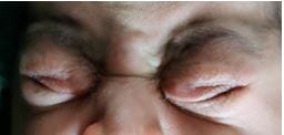
Fig 1: Clinical photograph of the neonate with congenital

opthal_241_02.JPG
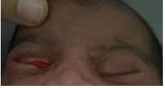
Fig 2: Clinical photograph of the neonate with congenital eyelid imbrication and floppy eyelid syndrome showing eyelid imbrication and floppy eyelid syndrome overlapping of the upper lid over the lower lid showing spontaneous eversion of the upper lid on gentle pulling of the skin of the upper lid.

opthal_241_03.JPG
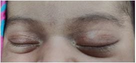
Figure 3: Clinical photograph of the neonate with congenital eyelid imbrication and floppy eyelid syndrome on day 5 showing the resolution.

Discussion

CEIS is identified by the overriding of the upper eyelid over the lower eyelid and is a malposition disorder of eyelids. Acquired imbrication eyelid syndrome is seen in adults above 40 years of age and in patients with lower lid


laxity and floppy eyelid syndrome who have undergone lateral tarsal strip procedures. CEIS is most commonly associated with FES.CEIS doesn’t require surgical intervention as it’s a self-limiting condition. Foreign body sensation caused by skin and eyelashes is relieved by instilling artificial tear substitutes. The association of Downs syndrome with FES has been noticed [2].

On systemic evaluation present case showed no features of Downs syndrome. Our case is different from previously reported due to the presence of moderate mechanical blepharoptosis which resolved within 5 days [1,3,4,5] [table .1].

During the early postnatal period, involutional changes and spontaneous tightening of canthal tendons lead to the resolution of blepharoptosis.

Table 1 Clinical presentation of reported cases of CEIS

Features Rumelts et al.[2] De Silva et al.[3] Odat et al.[1] Chandravanshi et al.[5] Present case
Age of presentation 3h 48h >24h 16h 5h
Sex Female Male Male Female female
Mode of delivery Vaginal Not mentioned Vaginal Caesarean section Caesarean section
Amount of overriding OU, 1.2mm OU, >1mm OD>OS; OS: 1-2mm OU 6mm OU,5mm
Canthi Laxed and longer Laxed Laxed Laxed and longer Laxed and longer
Spontaneous eversion of upper eyelids absent Present Present Present Present
Purulent discharge Not mentioned Not mentioned Present Present Absent
Recovery 1 week 2 months 3 weeks 1 week 5 days

The pathophysiology of combined CEIS and FES is laxity of the lateral canthal tendons and is similar to the pathophysiology of FES in adults. Spontaneous resolution of the floppy eyelid and eyelid imbrication is attributed to post-natal growth of the bony orbit.

All the cases have been under-reported as they have spontaneous natural resolution due to the apparent tightening of the canthal tendons.

CEIS though it resolves spontaneously identification of this condition is important to prevent complications like corneal epithelial defects and ulcers.


Conclusion

Congenital eyelid imbrication syndrome is a rare condition of uncertain cause and is self-limiting. In a neonate with congenital eyelid malposition, CEIS and CFES should be considered differential diagnoses. It is important to diagnose CEIS as it is associated with serious sight-threatening complications such as corneal epithelial defects and ulcers. Newborns having CEIS should be evaluated for FES. This case report helps in familiarizing ophthalmologists towards the identification of FES and CEIS.

Reference

01. Odat, Thabit Ali Abduallah Mustafa, and Janet Saeed Hina. Congenital combined eyelid imbrication and floppy eyelid syndrome. Journal of Optometry 3. 2 (2010): 91-93. [Crossref][PubMed][Google Scholar]

02. Laaribi N, Abdellaoui T, Houba A, Jeddou I, Habyebete S, Reda K, et al. Floppy eyelid et syndrome de Down : à propos d’un cas [Floppy eyelid and Down syndrome: Case report]. J Fr Ophtalmol. 2019 Apr;42(4):e173-e175. French. doi: 10.1016/j.jfo.2018.07.019 [Crossref][PubMed][Google Scholar]

03. Rumelt S, Kassif Y, Rehany U. Congenital eyelid imbrication syndrome. Am J Ophthalmol. 2004 Sep;138(3):499-501. doi: 10.1016/j.ajo.2004.04.023 [Crossref][PubMed][Google Scholar]

04. De Silva DJ, Fielder AR, Ramkissoon YD. Congenital eyelid imbrication syndrome. Eye (Lond). 2006 Sep;20(9):1103-4. doi: 10.1038/sj.eye.6702141 [Crossref][PubMed][Google Scholar]

05. Chandravanshi SL, Rathore MK, Tirkey ER. Congenital combined eyelid imbrication and floppy eyelid syndrome: case report and review of literature. Indian J Ophthalmol. 2013 Oct;61(10):593-6. doi: 10.4103/0301-4738.121086 [Crossref][PubMed][Google Scholar]