E-ISSN:2456-6454
P-ISSN:2581-4907
RNI:MPENG/2017/74152

Research Article

Fungal keratitis

Tropical Journal of Ophthalmology and Otolaryngology

2022 Volume 7 Number 5 September October
Publisherwww.medresearch.in

Multi-layered amniotic membrane transplantation for the management of corneal ulcers during the Covid pandemic

Joshi S.1*, Joshi H.2, Abhyankar A.3, Madan Deshpande C.4
DOI: https://doi.org/10.17511/jooo.2022.i05.01

1* Shilpa Joshi, Cornea Consultant & H.O.D, Medical Director Eye Bank, H.V. Desai Eye Hospital, Pune, Maharashtra, India.

2 Hema Joshi, Cornea Consultant, H.V. Desai Eye Hospital, Pune, Maharashtra, India.

3 Anannya Abhyankar, DNB Final year resident, H.V. Desai Eye Hospital, Pune, Maharashtra, India.

4 Col. Madan Deshpande, Chief Medical Director, H.V. Desai Eye Hospital, Pune, Maharashtra, India.

This study was done to assess the outcome of multilayer amniotic membrane transplantation (AMT) for severe progressive fungal keratitis & perforated corneal ulcers. With the COVID-19 pandemic-related lockdown and unavailability of donor corneas, multi-layered AMT was done in these cases. We hereby report nine such cases, all of which showed reduced inflammation, resolution of infiltrates, good epithelisation and no recurrence of infection. None needed TPK in subsequent weeks of follow-up. With palliative treatment of AMT, eyes with vision-threatening keratitis can be salvaged, till the availability of donor corneas.

Keywords: Multilayer amniotic membrane transplantation, COVID-19, Fungal keratitis

Corresponding Author How to Cite this Article To Browse
Shilpa Joshi, Cornea Consultant & H.O.D, Medical Director Eye Bank, H.V. Desai Eye Hospital, Pune, Maharashtra, India.
Email:
Shilpa Joshi, Hema Joshi, Anannya Abhyankar, Col. Madan Deshpande, Multi-layered amniotic membrane transplantation for the management of corneal ulcers during the Covid pandemic. Trop J Ophthalmol Otolaryngol. 2022;7(5):32-38.
Available From
https://opthalmology.medresearch.in/index.php/jooo/article/view/233

Manuscript Received Review Round 1 Review Round 2 Review Round 3 Accepted
2022-04-06 2022-04-08 2022-04-15 2022-04-22 2022-04-29
Conflict of Interest Funding Ethical Approval Plagiarism X-checker Note
Nil Nil Yes 18%

© 2022by Shilpa Joshi, Hema Joshi, Anannya Abhyankar, Col. Madan Deshpandeand Published by Siddharth Health Research and Social Welfare Society. This is an Open Access article licensed under a Creative Commons Attribution 4.0 International License https://creativecommons.org/licenses/by/4.0/ unported [CC BY 4.0].

Introduction

Fungal keratitis is the most common cause of infective keratitis and comprises at least 50% of all culture-positive cases in India.[1]. Various modalities of treatment are antifungal drops, Intrastromal injections of antifungal drugs for deep-seated ulcers, [2]. cyanoacrylate glue and a bandage contact lens for small perforations. [3]. In non-responding large ulcers, Therapeutic Keratoplasty (TPK) is currently considered the most effective method, for the elimination of infection & salvaging the eye.

With Coronavirus disease 2019 (COVID-19) spreading all across the globe, many countries, including India faced a lockdown. As per the Government notifications, all activities of the eye bank were halted and no donor corneas were available even for emergency procedures. In the past, many studies have proved that Amniotic membrane (AM), because of its special biological properties like lack of immunogenicity, promotion of epithelisation , inhibition of stromal inflammation , stabilisation of stromal thickness can help to heal corneal perforations and deep ulcers. [,4,5]. So we thought of treating cases of very progressive corneal ulcers with increasing infiltrates, ulcers approaching limbus, impending perforations and perforated ulcers, with multi-layered AM Transplantation (AMT). We present a series of nine cases which were salvaged with this technique.

Materials and Methods

This is a retrospective case series of very severe & progressive fungal ulcers, that presented to our cornea OPD from April 2020 – September 2020. All the cases, except one, had a history of vegetative trauma. All cases were previously given a mixed regimen of topical anti-bacterial & antifungals for a minimum of two weeks (one case) to two months (eight cases), before being referred to our hospital. The diagnosis was based on clinical features, smear and or culture of corneal scrapings. Three were positive for fungus on KOH. Two were large perforated ulcers with near total corneal melt & in one case Glue & BCL had been done. Of the remaining three cases, one posted perforation central ectatic cicatrix, the other was graft infection with impending perforation & third was of unknown etiology, with suspected endophthalmitis.


Technique

All cases were done by two cornea surgeons, (SJ, HJ) under peribulbar anaesthesia with supplementary intravenous sedation whenever required. Cryopreserved AM was washed with Balanced Salt Solution three times, to reduce any possibility of contamination.[6] . Excision of the infected cornea leaving behind a rim of the healthy noninfected cornea was done. Exudates on & behind the iris were removed carefully, sacrificing infiltrate-laden non-viable iris, if needed. A core vitrectomy was done if needed. The amniotic membrane was cut into appropriate, sized pieces &multiple layers (3-4) were placed onto the ulcer bed, with fibrin glue between each layer. Then a piece of AM was trimmed to adequately cover the ulcer bed, placed epithelial side up & sutured with 10-0 nylon interrupted sutures. Lastly, a large piece of AM was used to cover the entire cornea and secured with interrupted sutures placed (Sandwich Technique) and BCL was placed [7].

Postoperatively, patients were continued on topical antifungals, prophylactic antibiotics, antiglaucoma & tear substitute drops.

At each postoperative visit, the following were monitored:1) Epithelisation 2) Localisation of the infiltrates (location, size, depth) 3) Resolution of infection 4) Need for TPK

Results

Two cases (cases 1&5) with central corneal perforation & pseudocornea, healed well within 6-8 weeks, later after 3- 4 months, optical Penetrating Keratoplasty (P.K.) was done for them.

[Fig.1-3] with good visual recovery.

Technique

All cases were done by two cornea surgeons, (SJ, HJ) under peribulbar anaesthesia with supplementary intravenous sedation whenever required. Cryopreserved AM was washed with Balanced Salt Solution three times, to reduce any possibility of contamination.[6]. Excision of the infected cornea leaving behind a rim of the healthy noninfected cornea was done. Exudates on & behind the iris were removed carefully, sacrificing infiltrate-laden non-viable iris, if needed. A core vitrectomy was done if needed.


The amniotic membrane was cut into appropriate, sized pieces &multiple layers (3-4) were placed onto the ulcer bed, with fibrin glue between each layer. Then a piece of AM was trimmed to adequately cover the ulcer bed, placed epithelial side up & sutured with 10-0 nylon interrupted sutures.

Lastly, a large piece of AM was used to cover the entire cornea and secured with interrupted sutures placed (Sandwich Technique) and BCL was placed [7]. Postoperatively, patients were continued on topical antifungals, prophylactic antibiotics, antiglaucoma & tear substitute drops.

At each postoperative visit, the following were monitored:1) Epithelisation 2) Localisation of the infiltrates (location, size, depth) 3) Resolution of infection 4) Need for TPK

Figure 1 – Case 1

opthal_233_01.JPG

Figure 1A: Perforated fungal ulcer- Aspergillus

opthal_233_02.JPG

Figure 1B: Post-Multi-layered AMG

opthal_233_03.JPG
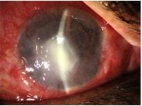
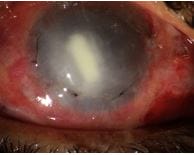
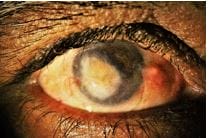
Figure 1C: Post healing- adherent leucoma
opthal_233_04.JPG
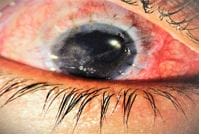
Figure 1D:
Post-Optical Keratoplasty

Figure 2 – Case 5
opthal_233_05.JPG
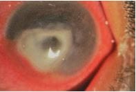
Figure 2A:Perforated fungal ulcer
opthal_233_06.JPG
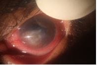
Figure 2B:
Post multi-layered AMG


opthal_233_07.JPG

Figure 2C:Post Optical PK

In four cases with large perforations & near total corneal melt, resolution of keratitis with globe integrity was achieved.

(Case 2,3,7,8) (Figures 3A and B)

opthal_233_08.JPG

Figure 3A:Perforated fungal ulcer with near-total corneal melt

opthal_233_09.JPG

Figure 3B: Post-AMG, healing of melt with irregular scarring


Table 1-Clinical findings & outcome of AMT in case series of 9 patients

Serial no Onset(weeks) Morphology of lesion Smear & culture Prior treatment Post AMG course
1 5 weeks Central corneal ulcer with deep stromal infiltrates, Hypopyon KOH- septate fungus CULTURE- Aspergillus 6 intrastromal voriconazole injections Resolved with corneal scarring, optical P.K.done later
2 6 weeks Perforated corneal ulcer with near-total corneal melt KOH- septate fungus Topical antifungals& antibiotics Epithelisation of the lesion, no reinfection, restored anatomical integrity
3 18 days corneal abscess with impending perforation, 360◦ choroidal KOH- septate fungus Topical antibiotics & antifungals Epithelisation, resolution of ulcer
4 7 weeks Deep stromal ulcer with endoexudates, suspected endophthalmitis KOH negative Intracameral & intravitreal ceftazidime & voriconazole injections Epithelisation, resolution of endophthalmitis, no reinfection, Phthisical eye
5 2 weeks Central ulcer with impending perforation KOH & Culture negative 3 intrastromal voriconazole injections Resolved keratitis with corneal scarring.Optical P.K.done later
6 5 weeks Central progressive ulcer Not done Glue BCL did (outside hospital) Healed with anterior staphyloma formation. Tectonic P.K. done
7 3 weeks Total corneal melt with IOL extruding out Not done Topical antibiotics antifungals 1 week FU- beginning of epithelistion Lost to FU
8 8 weeks Central ectatic cicatrix with iris incarceration Not done Topical antibiotics Corneal scarring
9 6 weeks Failed graft with decematocele Not done P.K. done twice (outside hospital), Glue BCL done Resolution of infection.


Figure 4 – Case 6

opthal_233_10.JPG
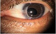
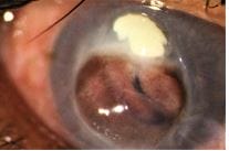
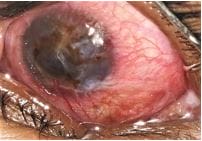
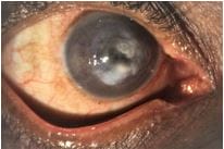
Figure 4A:Corneal ulcer with large descemetoceleopthal_233_11.JPG
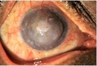
Figure 4B:Post-AMG healed ulcer with anterior staphyloma

One case healed with anterior staphyloma formation later required tectonic P.K. (Case 6) (Figure 4A and B)

In one case with perforation & endophthalmitis, the progression of infection was prevented, but the eye became pre phthisical. Case with graft infection also healed well post AMT, was advised repeat optical PK but refused surgery. The findings of patients are summarized in Table 1. 

Discussion

During the COVID-19 pandemic, due to the stoppage of the Hospital Cornea Retrieval Program (HRCP), there was an acute shortage of donor corneas. Hence we resorted to, multilayer AMT for patients with severe keratitis with large perforations. Amniotic membrane (AM), produces various proteinase inhibitors and the stromal matrix has various growth factors like TGF ß which contribute to its anti-inflammatory, anti-angiogenic, anti-scarring & pro-regenerative abilities.[4].

AM can reduce tissue damage & promote wound healing. [4]. Although in the past there were concerns about the recurrence of infection with AM transplantation, recent reports show that cryopreserved AM with intrinsic anti-infective properties has been used successfully in the treatment of non-healing corneal ulcers. [7,8]. It has been used in non-healing bacterial ulcers, [9]. also in acanthamoeba & HSV necrotising stromal keratitis.[10,5]. It has been used in Aspergillus, Fusarium & other persistent fungal infections [10,11]. with very low and treatable recurrence of infection even in active fungal keratitis [11]. In our case series too, there was no reinfection. AM is used as a single-layer inlay or overlay, multi-layered inlay or sandwich technique depending on the depth of the ulcer. [7,10].

Add what new this study adds to existing knowledge under the separate heading.

Newer Insights: In many cases reporting the use of AMG for nonhealing ulcers, keratitis was in the form of persistent epithelial defect with early sterile stromal melt.in these cases, a temporary AM as a patch, either sutured or self-retained (PROKERA ) resulted in surface healing [7,10]. In contrast, all our cases were infective melts or perforations. So a single-layer AM graft would have melted or dissolved due to overwhelming infection & inflammation. There are reports of the use of AMT for frank infections too, but those were central or paracentral small and superficial ulcers.[8,10]. unlike our series where all cases were severe progressive, limbus reaching or perforated ulcers. AMT has been used quite effectively for bacterial, acanthamoeba & herpes simplex keratitis, [5,7]. but in very few fungal keratitis cases, unlike our series where the majority were fungal. Another difference in the technique of AMG is most case series report the use of a single-layer overlay, or multi-layered inlay technique [8,11,12]. whereas we have used a combination or sandwich technique and method of using several pieces of AM as an inlay with the use of fibrin glue &onlay patch with sutures probably worked like a tectonic patch graft.

As all our cases were very severe keratitis, a single layer of AM would have melted within a few days. The use of multi-layered AM in ulcers with marginal residual stromal tissue, prevented aqueous leak & anterior chamber collapse, whereas, in perforated ulcers, it provided some tectonic support, possibly


by restoring corneal integrity & healing by scar formation [12].

Alternatively, conjunctival flap (CF) has been used by Gunderson& others [13]. in the past for the treatment of chronic ulcers. However, it might not work for large perforated ulcers due to difficulty in dissection, mobilisation, suturing with possible complications, buttonholing, and retraction of the flap & corneal perforation [14].

Conclusion

In our small case series, we achieved the elimination of infection with the restoration of globe integrity. we found that AMT is effective in the resolution of ulcers with the preservation of globe integrity & salvaging the eye when the donor cornea is unavailable. So we recommend the use of cryopreserved AMT in infectious keratitis cases not responding to medical management. It is definitely worth using this technique of multi-layered AMG, to buy time, till donor cornea becomes available. For very large perforations, conjunctival or Tennon’s graft may not suffice, so AMT becomes a globe-saving surgery in such cases.

Authors’ Contribution: Shilpa Joshi performed many cases of these series, maintained proper documentation of follow-up visits, clinical photos etc., helped in compiling the data, wrote the manuscript & revised it many times. Hema Joshi operated on some cases of these series. Ananya Abhyankar helped in compiling the data and in writing the initial draft of the manuscript. Col Deshpande gave some valuable inputs to improve the quality of the manuscript.

Reference

01. Thomas PA. Current perspectives on ophthalmic mycoses. Clin Microbiol Rev. 2003 Oct;16(4):730-97. doi: 10.1128/CMR.16.4.730-797.2003 [Crossref][PubMed][Google Scholar]

02. Sharma N, Agarwal P, Sinha R, Titiyal JS, Velpandian T, Vajpayee RB. Evaluation of intrastromal voriconazole injection in recalcitrant deep fungal keratitis: case series. Br J Ophthalmol. 2011 Dec;95(12):1735-7. doi: 10.1136/bjo.2010.192815 [Crossref][PubMed][Google Scholar]

03. Garg P, Gopinathan U, Nutheti R, Rao GN.

Clinical experience with N-butyl cyanoacrylate tissue adhesive in fungal keratitis. Cornea. 2003 Jul;22(5):405-8. doi: 10.1097/00003226-200307000-00003 [Crossref][PubMed][Google Scholar]

04. Dua HS, Gomes JA, King AJ, Maharajan VS. The amniotic membrane in ophthalmology. Surv Ophthalmol. 2004 Jan-Feb;49(1):51-77. doi: 10.1016/j.survophthal.2003.10.004 [Crossref][PubMed][Google Scholar]

05. Heiligenhaus A, Li H, Hernandez Galindo EE, Koch JM, Steuhl KP, Meller D. Management of acute ulcerative and necrotising herpes simplex and zoster keratitis with amniotic membrane transplantation. Br J Ophthalmol. 2003 Oct;87(10):1215-9. doi: 10.1136/bjo.87.10.1215 [Crossref][PubMed][Google Scholar]

06. Marsit NM, Sidney LE, Britchford ER, McIntosh OD, Allen CL, Ashraf W, et al. Validation and assessment of an antibiotic-based, aseptic decontamination manufacturing protocol for therapeutic, vacuum-dried human amniotic membrane. Sci Rep. 2019 Sep 6;9(1):12854. doi: 10.1038/s41598-019-49314-7 [Crossref][PubMed][Google Scholar]

07. Yin HY, Cheng AMS, Tighe S, Kurochkin P, Nord J, Dhanireddy S, et al. Self-retained cryopreserved amniotic membrane for treating severe corneal ulcers: a comparative, retrospective control study. Sci Rep. 2020 Oct 12;10(1):17008. doi: 10.1038/s41598-020-73672-2 [Crossref][PubMed][Google Scholar]

08. Liu J, Li L, Li X. Effectiveness of Cryopreserved Amniotic Membrane Transplantation in Corneal Ulceration: A Meta-Analysis. Cornea. 2019 Apr;38(4):454-462. doi: 10.1097/ICO.0000000000001866 [Crossref][PubMed][Google Scholar]

09. Sangwan VS, Burman S, Tejwani S, Mahesh SP, Murthy R. Amniotic membrane transplantation: a review of current indications in the management of ophthalmic disorders. Indian J Ophthalmol. 2007 Jul-Aug;55(4):251-60. doi: 10.4103/0301-4738.33036 [Crossref][PubMed][Google Scholar]

10. Mohan S, Budhiraja I, Saxena A, Khan P, Sachan SK. Role of multilayered amniotic membrane transplantation for the treatment of resistant corneal ulcers in North


India. Int Ophthalmol. 2014 Jun;34(3):485-91. doi: 10.1007/s10792-013-9834-3 [Crossref][PubMed][Google Scholar]

11. Kim JS, Kim JC, Hahn TW, Park WC. Amniotic membrane transplantation in infectious corneal ulcer. Cornea. 2001 Oct;20(7):720-6. doi: 10.1097/00003226-200110000-00010 [Crossref][PubMed][Google Scholar]

12. Chen HC, Tan HY, Hsiao CH, Huang SC, Lin KK, Ma DH. Amniotic membrane transplantation for persistent corneal ulcers and perforations in acute fungal keratitis. Cornea. 2006 Jun;25(5):564-72. doi: 10.1097/01.ico.0000227885.19124.6f [Crossref][PubMed][Google Scholar]

13. Berguiga M, Mameletzi E, Nicolas M, Rivier D, Majo F. Long-term follow-up of multilayer amniotic membrane transplantation (MLAMT) for non-traumatic corneal perforations or deep ulcers with descemetocele. Klin Monbl Augenheilkd. 2013 Apr;230(4):413-8. doi: 10.1055/s-0032-1328394 [Crossref][PubMed][Google Scholar]

14. Alino AM, Perry HD, Kanellopoulos AJ, Donnenfeld ED, Rahn EK. Conjunctival flaps. Ophthalmology. 1998 Jun;105(6):1120-3. doi: 10.1016/S0161-6420(98)96017-1 [Crossref][PubMed][Google Scholar]

15. Zhong J, Wang B, Li S, Deng Y, Huang H, Chen L, et al. Full-thickness conjunctival flap covering surgery combined with amniotic membrane transplantation for severe fungal keratitis. Exp Ther Med. 2018 Mar;15(3):2711-2718. doi: 10.3892/etm.2018.5765 [Crossref][PubMed][Google Scholar]