E-ISSN:2456-6454
P-ISSN:2581-4907
RNI:MPENG/2017/74152

Research Article

Tympanoplasty

Tropical Journal of Ophthalmology and Otolaryngology

2021 Volume 6 Number 6 November December
Publisherwww.medresearch.in

Comparative Study of Clinical Outcome between Microscopic and Endoscopic Type I Tympanoplasty

Yadav M.1*, Hussain R.2, Monga J.3, Naik S.4
DOI: https://doi.org/10.17511/jooo.2021.i06.03

1* Mayank Yadav, Assistant Professor, Department of Otorhinolaryngology, SHKM GMC, Nuh, Haryana, India. ORCID: 0000-0002-9729-752X

2 Raj Tajamul Hussain, Senior Registrar, Department of Otorhinolaryngology, SHKM GMC, Nuh, Haryana, India. ORCID: 0000-0002-9795-3697

3 Jasdeep Monga, Associate professor, Department of Otorhinolaryngology, SHKM GMC, Nuh, Haryana, India. ORCID: 0000-0001-5764-063X

4 S.M. Naik, Professor, Department of Otorhinolaryngology, SHKM GMC, Nuh, Haryana, India. ORCID: 0000-0002-7159-5019

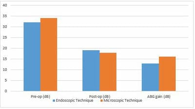
Introduction: Tympanoplasty is one of the most common surgical procedures in ENT. It can be performed with the help of a microscope or endoscope. We carried out a prospective randomized comparative study in our tertiary care centre to evaluate and compare the results of endoscopic and conventional microscopic tympanoplasty in terms of hearing outcome, surgical time and graft success rate. Materials and Methods: This prospective randomized comparative study was conducted in the Department of Otorhinolaryngology, SHKM Hospital, an associated hospital of GMC Nuh, Haryana, India, for 12 months from October 2019 to September 2020. Eighty patients with central perforation were randomly divided into two equal groups of 40 patients each. In the first group, an endoscope was used, and in the second group, the microscope was used to do type 1 tympanoplasty. The patients were kept on follow-up for six months. The graft success rates, hearing outcomes and duration of surgery in patients were compared in both groups. Results: Graft success rates were 90% and 92.5% for the endoscopic and microscopic groups, respectively (p > 0.05). Postoperative air-bone gap values significantly improved in both groups (p < 0.05). The average duration of surgery was significantly shorter in the endoscopic group (mean 42.9 min) relative to the microscopic group (mean 57.2 min) (p < 0.05). Conclusion: In managing chronic otitis media, endoscopic transcanal tympanoplasty can be a viable alternative to conventional microscopic tympanoplasty, with comparable graft success rates and hearing outcomes.

Keywords: CSOM. Endoscopic tympanoplasty. Microscopic tympanoplasty, Perforation, Operative duration, ABG

Corresponding Author How to Cite this Article To Browse
Mayank Yadav, Assistant Professor, Department of Otorhinolaryngology, SHKM GMC, Nuh, Haryana, India.
Email:
Mayank Yadav, Raj Tajamul Hussain, Jasdeep Monga, S.M. Naik, Comparative Study of Clinical Outcome between Microscopic and Endoscopic Type I Tympanoplasty. Trop J Ophthalmol Otolaryngol. 2021;6(6):121-127.
Available From
https://opthalmology.medresearch.in/index.php/jooo/article/view/226

Manuscript Received Review Round 1 Review Round 2 Review Round 3 Accepted
2021-12-04 2021-12-06 2021-12-13 2021-12-20 2021-12-27
Conflict of Interest Funding Ethical Approval Plagiarism X-checker Note
Nil Nil Yes 16%

© 2021by Mayank Yadav, Raj Tajamul Hussain, Jasdeep Monga, S.M. Naikand Published by Siddharth Health Research and Social Welfare Society. This is an Open Access article licensed under a Creative Commons Attribution 4.0 International License https://creativecommons.org/licenses/by/4.0/ unported [CC BY 4.0].

Introduction

Chronic otitis media is any structural change in the middle ear system associated with a permanent defect in the tympanic membrane (TM) [1]. The goal of tympanoplasty is to eradicate the disease from the middle ear and restore the hearing mechanism. Wullstein divided tympanoplasty into five types in 1956. When the middle ear ossicles are intact, type I tympanoplasty involves the repair of the perforated tympanic membrane. It can be done via endaural, postural, or transcanal routes, and the graft can be placed employing overlay, interlay, or underlay techniques. Incorrect and inadequate treatment of chronic otitis media (COM) may cause severe complications [2]. Conventional microscopic tympanoplasty with a postauricular incision remains the most effective and commonly used treatment for chronic otitis media, particularly in cases of anterior or large tympanic membrane perforation with anterior bony overhang [3]. Endoscopes were first used in middle ear surgery in the 1990s and have since been accepted worldwide [4, 5]. Endoscopes are currently employed as a primary or auxiliary device in nearly all types of middle ear surgeries, including chronic otitis surgery, stapes surgery, and cochlear implantation [6,7]. Endoscopy is commonly used in tympanoplasty because it gives a clear surgical field and allows for exposure of the circumferential TM, and avoids a postauricular incision and subsequent scar formation. Unlike a microscope, the endoscope is easily transportable and ideal for in-ear surgery camps. Despite these benefits, the endoscopic approach has its drawbacks. The endoscope lacks adequate microscopic magnification and focus; also, this procedure necessitates the surgeon operating one-handed, holding the endoscope in one hand and other equipment or suction in the freehand. Instruments may crowd the ear canal during the endoscopic approach, and the endoscope may get frequently or substantially coated with blood [8]. Endoscopic surgery provides 2D pictures, and 2D images lack depth awareness [9], making it difficult to raise the graft to establish touch with the perforation's edge. However, advanced high-definition camera systems can provide considerably more delicate endoscopic views with improved contrast. There is a shortage of research on endoscopic tympanoplasty.

Furthermore, there have

been few comparative studies between standard microscopic techniques and endoscopic procedures [10]. As a result, a comparative evaluation of the endoscopic and microscopic techniques of tympanoplasty is warranted. This study aims to evaluate and compare the results of endoscopic and conventional microscopic tympanoplasty in terms of hearing outcome, surgical time and graft success rate. The aim was to compare the clinical benefits of permeatal endoscopic tympanoplasty to conventional postaural microscopic surgery.

Material and Methods

This prospective randomized comparative study was conducted in the Department of Otorhinolaryngology, SHKM Hospital, an associated hospital of GMC Nuh, Haryana, India, for 12 months from October 2019 to September 2020. The hospital's institutional review board approved this study. All patients who participated in our study provided written informed consent. A total of 80 patients with central tympanic membrane perforation were recruited. Patients who fulfilled all of the inclusion and exclusion criteria and were ready to provide their voluntary consent for study participation were divided into two groups depending on the surgical intervention they underwent. Group 1 underwent endoscopic tympanoplasty (40 ears from 40 patients), while Group 2 underwent microscopic tympanoplasty (40 ears from 40 patients). Under local anesthesia, all patients underwent type 1 tympanoplasty, and the underlay technique with temporalis fascia as graft material was employed across all cases. A single surgeon operated on all of the cases. The pre-and postoperative audiograms, graft uptake, and surgery time were compared in both groups.

Inclusion criteria

  1. Chronic suppurative otitis media (CSOM) with dry central perforation
  2. Age >18years
  3. Dry ear for at least last six months
  4. Patients with conductive hearing loss < 40 dB

Exclusion criteria

  1. Active ear discharge
  2. Cholesteatoma and granulation tissue in the middle ear
  3. CSOM with ossicular chain defects

  1. Postoperative residual and recurring tympanic membrane perforations

Surgical equipment: The procedure was performed with a 0-degree 4 mm rigid endoscope. After the tympanomeatal flap was elevated, a 30 degree 4 mm rigid endoscope was employed for some time in the middle of the surgery to inspect the middle ear cavity. Moller-Wedel surgical microscope (GmbH Rosergarten Germany) was used for microscopic tympanoplasty. The display and video system were from Karl Storz. The surgical instruments were conventional micro-instruments used in otology.

Surgical Technique: All the patients were operated on under local anesthesia. Intramuscular injections of 1 ampoule for twin and one ampoule Phenergan were administered as a premedication. The external auditory canal was anaesthetized with 2% lignocaine with 1 in 100,000 adrenaline injections. Under local anesthesia, a temporalis fascia graft was harvested and allowed to dry.

Endoscopic surgery: Cerumen and external auditory canal (EAC) secretions were removed before surgery. The EAC's long hair was cropped. Using the 0 degrees 4 mm endoscope, the perforation margin was freshened with a sickle knife or an angled pick. A curvilinear incision was made about 5 mm lateral to the annulus, extending between 11 o'clock and 1 o'clock position, with a round knife. The skin was gradually elevated away from the external canal's bone. The annulus was upgraded, and the middle ear mucosa was incised. The tympanomeatal flap was then elevated from the handle of the malleus to gain access to the middle ear cavity. After skeletonizing the malleus handle, the temporalis fascia graft was placed using the underlay technique. The tympanomeatal flap protected the temporalis fascia graft. Gelfoam pieces were put over the tympanomeatal flap.

Microsurgery: Cerumen and EAC secretions were cleaned before surgery. The EAC's long hair was cropped.  The affected ear was adequately positioned by turning the patient's head towards the opposite side. The external auditory canal was exposed via Wilde's incision. After anteriorly retracting the pinna with a Mollison's retractor, the perforation margin was freshened with a sickle knife. The tympanomeatal flap was raised from 5 mm lateral to the tympanic annulus, skeletonizing the malleus handle. The temporalis fascia graft

was spread uniformly below the handle of the malleus. The tympanomeatal flap was repositioned over the temporalis fascia. Gelfoam pieces were placed over the tympanomeatal flap. The postural wound was sutured with 3-0 silk and stitches removed after one week. The length of the surgery was also documented. On the second postoperative day, all patients were discharged with oral antibiotics and analgesics for seven days.  The aural wick was removed after one week.

Follow-up: Postoperative follow-up evaluations included pure tone audiometry, tympanometry, and endoscopic or microscopic evaluation of the graft condition at 1, 3, and 6 months. The final audiometric assessment done at six months was taken for analysis in our study. Averages of 0.5, 1.0, and 2.0 kHz were used to assess hearing thresholds, including air conduction and bone conduction thresholds. In each examination, the air-bone gap (ABG) was also estimated.

Statistical analyses: The T-test and chi-square test were calculated using Statistical Package for the Social Sciences (SPSS) software (IBM Corporation; NY, USA). A p-value < 0.05 was considered to be statistically significant.

Results

The mean age was 25.3 years (range 19—61 years) in group I, with 19 (47.5%) males and 21 (52.5%) females, while the average age was 33.5 years in Group II, with 26 (65%) males and 14 (35%) females (range 19—72 years). Endoscopic type 1 tympanoplasty was performed on 29 (72.5%) right and 11 (27.5%) left ears in Group I. In the microscopic tympanoplasty group, 22 (55%) of the ears operated on were right, and 18 (45%) were left.

There was no previous history of ear surgery in either group. All patients were discharged on 2nd postoperative day. Table 1 shows the demographic characteristics and surgical results of subjects undergoing endoscopic and microscopic type 1 tympanoplasty.

After the first month following surgery, 32 patients out of 40 in the endoscopic group had intact tympanic membrane, and by the third month, 36 patients out of 40 had intact tympanic membrane. Thirty-six patients had intact tympanic membrane at the end of the 6-month


follow-up period. All five graft failure patients had developed a postoperative infection and were erratic in their follow-up.

After one month, 33 patients out of 40 in the microscopic group had intact tympanic membrane, and by the third month, 37 patients out of 40 had intact tympanic membrane. Thirty-seven patients had intact tympanic membrane at the end of the 6-month follow-up. All four graft failure patients had developed a postoperative infection and were not following up regularly.

In conclusion, after six months of follow-up, the graft success rates for Group I (36 out of 40 ears) and Group II (37 out of 40 ears) were 90% and 92.5%, respectively. There was no significant difference in graft success rates between the groups (p > 0.05). For patients in Groups I and II, the mean operative time (SD) was 42.9 ± 10.1 minutes (range 30—56 minutes) and 57.2 ± 4.9 minutes (range 46—75 minutes), respectively.  The duration of the procedure in group I was considerably shorter than in Group II (p < 0.05).

Table 1: Demographic characteristics and surgical outcomes of the patients. 

  Endoscopic Group (n=40) Microscopic Group (n=40) p-value*
Age (mean age) 25.3 (19-61) 33.5 (19-72)  
Gender  
Male 19 (47.5%) 26 (65%)
Female 21 (52.5%) 14 (35%)
Side  
Right 29 (72.5%) 22 (55%)
Left 11 (27.5%) 18 (45%)
Graft uptake rate (%) 90% 92.5% >0.05
Mean operative duration (mean ± SD) 42.9 ± 10.1 57.2 ± 4.9 <0.05

SD, standard deviation.

*A p-value less than 0.05 was considered to be statistically significant.

The preoperative air-bone gap in the endoscopic group was 30–35 dB in 28 patients and 35–40 dB in 12 patients (Fig. 1) with a mean pure tone Air-Bone gap of 32.17 dB pre-operatively. At the end of a 6-month follow-up, all 36 cases of graft uptake in this group had a mean pure tone Air-Bone gap of 19.21 dB. 

The preoperative air-bone gap in the microscopic group was 30–35 dB in 23 patients and 35–40 dB in 17 patients (Fig. 1) with a mean pure tone Air-Bone gap of 34.21 dB pre-operatively. At the end of the 6-month follow-up, all 37 graft uptake patients had a mean pure tone Air-Bone gap of 18 dB. 

In conclusion, in our research, the postoperative air-bone gap was below 20 dB in 90% of endoscopic group patients and 92.5% of microscopic group patients. The mean ABG gain in the endoscopic group was 12.96 dB (SD = 3.88) and 16.21 dB (SD = 4.71) in the microscopic group (Fig. 4). This finding was statistically significant (p-value < 0.05).

opthal_226_01.JPG

Figure 1: Pre- and postoperative air-bone gap values in both endoscopic and microscopic techniques.

Discussion

The main goals of chronic otitis media treatment are to repair the tympanic membrane, eradicate the chronic infection, and, if necessary, restore ossicular chain integrity and mobility by ossiculoplasty or artificial prosthesis [4, 5, 11, 12]. Various surgical procedures, artificial prosthesis, graft materials, and grafting techniques have been used [6, 7]. El-Guindy [13] was the first to publish a series on endoscopic myringoplasty in 1992. However, Thomassin et al. [14] were the first to report the use of endoscopic assisted surgery to look at hidden areas during cholesteatoma surgery. Stereo vision and bimanual handling are the significant benefits of the microscopic technique. Despite providing direct exposure, microscopes require constant adjustment and maybe ineffective when dealing with protruding structures, especially the anterior wall. The endoscopic technique offers a new perspective to understanding anatomy, pathology, and operative techniques.


A surgeon can do minimally invasive procedures while protecting the anatomy with a thin, rigid endoscope, enabling functional reconstruction during surgery [15].

Furukawa et al. [16] found that in 12% of cases, the circumference of the perforation could not be examined using a microscope before denuding. Furthermore, after refreshing the margins, the whole perforation was not visible in 20% of cases. Endoscopy, on the other hand, can reveal the entire tympanic membrane in one field with clear visualization of the perforation margins, even if the ear canal is small or protruding. When evaluating the success of perforation closure with endoscopic and microscopic type 1 tympanoplasty, some authors had superior outcomes with the microscope [10,17-19] and others with the endoscope [20,21], although these differences were statistically insignificant.

Pap et al. [22] conducted a meta-analysis of endoscopic versus microscopic type I tympanoplasties and discovered that endoscopic surgical outcomes were comparable to microscopic type I tympanoplasty. In our study, the success rates of the microscopic approach for perforation repair and ABG closure were just marginally higher.

According to Tseng et al., graft success rates for endoscopic and microscopic tympanoplasty were 85.1% and 86.4%, respectively, with no significant difference [23]. The graft success rates in the endoscopic and microscopic techniques were 83.3% --100% and 82.4% --100%, respectively, in the studies comparing the endoscopic and microscopic tympanoplasty [24-27]. The current study is consistent with the existing literature in that there was no significant difference in graft success rates between the endoscopic and microscopic groups.

In studies comparing endoscopic and microscopic Type 1 tympanoplasty, the pre-and postoperative ABG values were between 28.5--46.4 dB and 18.1--8.1 dB, respectively. The current studies found that postoperative ABG values improved significantly in either endoscopic or microscopic tympanoplasty [19, 25-28]. In our study, postoperative ABG values improved significantly in both groups compared to preoperative ABG values. Furthermore, we found no statistically significant difference between Groups I and II in postoperative ABG gain. Endoscopic tympanoplasty is a safe and reasonable alternative to the conventional microscopic retroauricular

approach in Type 1 tympanoplasty in terms of hearing outcomes.           

The advantages of the endoscopic technique also include a reduction in operative time, which leads to a reduction in anesthesia duration and accompanying side effects, as well as a lesser burden on the surgeon's concentration. Ghaffar et al. [29] found that the average operation time for 34 patients undergoing endoscopic tympanoplasty was 62.85 minutes. The mean operating time in our study was 42.9 ± 10.1 minutes for the 40 ears that had the endoscopic technique, compared to 57.2 ± 4.9 minutes for the microscopic approach on a similar number of ears, which is a statistically significant difference (p-value < 0.05).  The preparation of the microscope, as well as the time required to harvest the graft and adjust the microscope, were the key variables accountable for this discrepancy in our hospital. 

In conclusion, the endoscopic approach for tympanoplasty offers superior visualization and shorter operative times than conventional surgery, in addition to equal hearing outcomes and perforation rates. Other advantages of this surgical technique include a lower rate of tissue injury, better cosmetic outcomes, and lower rates of perioperative nausea and vomiting.

Conclusion

Endoscopic tympanoplasty is the way of the future. With outcomes comparable to microscopic techniques and other advantages, the endoscope may surpass the microscope in popularity. Endoscopes may be used in the future for all types of ear procedures such as tympanoplasty, stapedotomy, cholesteatoma surgery, and cochlear implant. We advocate using an endoscope for type I tympanoplasties, either as a primary tool or as an adjunct tool to the microscope, due to the enhanced imaging via a minimal access approach and comparable results to microscopic surgery.

What does the study add to existing knowledge?  Our study substantiates the new emerging trend of endoscope utilization in performing ear surgeries and found results of endoscopic tympanoplasties on par with the current standard microscopic tympanoplasties.

Contribution by authors: Author 1: (Yadav M) conceived the project proposal,


manuscript writing and performed surgeries. Author 2: (Tajamul Hussain R) contributed in surgical work. Author 3: (Monga J) contributed in result compilation and analysis. Author 4 (Naik SM) contributed in final proof reading of the manuscript.

Reference

01. Bedajit RK, Mallik P, Kumar P, Devi HP, Sudhiranjan T, Singh OP et al. Endoscopic transcanal mastoidectomy with tympanoplasty. Natl J Otorhinolaryngol Head Neck Surg 3. 12 (2015): 10-11. [Crossref][PubMed][Google Scholar]

02. Morris PS, Leach AJ. Acute and chronic otitis media. Pediatr Clin North Am. 2009 Dec;56(6):1383-99. doi: 10.1016/j.pcl.2009.09.007 [Crossref][PubMed][Google Scholar]

03. Rizer FM. Overlay versus underlay tympanoplasty. Part II: the study. Laryngoscope. 1997 Dec;107(12 Pt 2):26-36. doi: 10.1097/00005537-199712001-00002 [Crossref][PubMed][Google Scholar]

04. Tarabichi M. Endoscopic middle ear surgery. Ann Otol Rhinol Laryngol. 1999 Jan;108(1):39-46. doi: 10.1177/000348949910800106 [Crossref][PubMed][Google Scholar]

05. Panetti G, Cavaliere M, Panetti M, Marino A, Iemma M. Endoscopic tympanoplasty in the treatment of chronic otitis media: our experience. Acta Otolaryngol. 2017 Mar;137(3):225-228. doi: 10.1080/00016489.2016.1227475 [Crossref][PubMed][Google Scholar]

06. Yiannakis CP, Sproat R, Iyer A. Preliminary outcomes of endoscopic middle-ear surgery in 103 cases: a UK experience. J Laryngol Otol. 2018 Jun;132(6):493-496. doi: 10.1017/S0022215118000695 [Crossref][PubMed][Google Scholar]

07. Güneri EA, Olgun Y. Endoscope-Assisted Cochlear Implantation. Clin Exp Otorhinolaryngol. 2018 Jun;11(2):89-95. doi: 10.21053/ceo.2017.00927 [Crossref][PubMed][Google Scholar]

08. Yadav SP, Aggarwal N, Julaha M, Goel A. Endoscope-assisted myringoplasty. Singapore Med J. 2009 May;50(5):510-2. [Crossref][PubMed][Google Scholar]

09. Mohindra S, Panda NK. Ear surgery without microscope; is it possible. Indian J Otolaryngol Head Neck Surg. 2010 Jun;62(2):138-41. doi: 10.1007/s12070-010-0033-5 [Crossref][PubMed][Google Scholar]

10. Harugop AS, Mudhol RS, Godhi RA. A comparative study of endoscope assisted myringoplasty and micrsoscope assisted myringoplasty. Indian J Otolaryngol Head Neck Surg. 2008 Dec;60(4):298-302. doi: 10.1007/s12070-008-0099-5 [Crossref][PubMed][Google Scholar]

11. House Wf, Sheehy JL. Myringoplasty. Use of ear canal skin compared with other techniques. Arch Otolaryngol. 1961 Apr;73:407-15. doi: 10.1001/archotol.1961.00740020417009 [Crossref][PubMed][Google Scholar]

12. Kartush JM, Michaelides EM, Becvarovski Z, LaRouere MJ. Over-under tympanoplasty. Laryngoscope. 2002 May;112(5):802-7. doi: 10.1097/00005537-200205000-00007 [Crossref][PubMed][Google Scholar]

13. el-Guindy A. Endoscopic transcanal myringoplasty. J Laryngol Otol. 1992 Jun;106(6):493-5. doi: 10.1017/s0022215100119966 [Crossref][PubMed][Google Scholar]

14. Thomassin JM, Korchia D, Doris JM. Endoscopic-guided otosurgery in the prevention of residual cholesteatomas. Laryngoscope. 1993 Aug;103(8):939-43. doi: 10.1288/00005537-199308000-00021 [Crossref][PubMed][Google Scholar]

15. Aoki K. Advantages of endoscopically assisted surgery for attic cholesteatoma. Diagn Ther Endosc. 2001;7(3-4):99-107. doi: 10.1155/DTE.7.99 [Crossref][PubMed][Google Scholar]

16. Furukawa T, Watanabe T, Ito T, Kubota T, Kakehata S. Feasibility and advantages of transcanal endoscopic myringoplasty. Otol Neurotol. 2014 Apr;35(4):e140-5. doi: 10.1097/MAO.0000000000000298 [Crossref][PubMed][Google Scholar]

17. Jyothi AC, Shrikrishna BH, Kulkarni NH, Kumar A. Endoscopic Myringoplasty Versus Microscopic Myringoplasty in Tubotympanic CSOM: A Comparative Study of


120 Cases. Indian J Otolaryngol Head Neck Surg. 2017 Sep;69(3):357-362. doi: 10.1007/s12070-017-1147-9 [Crossref][PubMed][Google Scholar]

18. Maran RK, Jain AK, Haripriya GR, Jain S. Microscopic Versus Endoscopic Myringoplasty: A comparative study. Indian J Otolaryngol Head Neck Surg. 2019 Nov;71(Suppl 2):1287-1291. doi: 10.1007/s12070-018-1341-4 [Crossref][PubMed][Google Scholar]

19. Dündar R, Kulduk E, Soy FK, Aslan M, Hanci D, Muluk NB, Cingi C. Endoscopic versus microscopic approach to type 1 tympanoplasty in children. Int J Pediatr Otorhinolaryngol. 2014 Jul;78(7):1084-9. doi: 10.1016/j.ijporl.2014.04.013 [Crossref][PubMed][Google Scholar]

20. Plodpai Y, Paje N. The outcomes of overlay myringoplasty: Endoscopic versus microscopic approach. Am J Otolaryngol. 2017 Sep-Oct;38(5):542-546. doi: 10.1016/j.amjoto.2017.05.007 [Crossref][PubMed][Google Scholar]

21. Ohki M, Kikuchi S, Tanaka S. Endoscopic Type 1 Tympanoplasty in Chronic Otitis Media: Comparative Study with a Postauricular Microscopic Approach. Otolaryngol Head Neck Surg. 2019 Aug;161(2):315-323. doi: 10.1177/0194599819838778 [Crossref][PubMed][Google Scholar]

22. Pap I, Tóth I, Gede N, Hegyi P, Szakács Z, Koukkoullis A, et al. Endoscopic type I tympanoplasty is as effective as microscopic type I tympanoplasty but less invasive-A meta-analysis. Clin Otolaryngol. 2019 Nov;44(6):942-953. doi: 10.1111/coa.13407 [Crossref][PubMed][Google Scholar]

23. Tseng CC, Lai MT, Wu CC, Yuan SP, Ding YF. Comparison of the efficacy of endoscopic tympanoplasty and microscopic tympanoplasty: A systematic review and meta-analysis. Laryngoscope. 2017 Aug;127(8):1890-1896. doi: 10.1002/lary.26379 [Crossref][PubMed][Google Scholar]

24. Kuo CH, Wu HM. Comparison of endoscopic and microscopic tympanoplasty. Eur Arch Otorhinolaryngol. 2017 Jul;274(7):2727-2732. doi: 10.1007/s00405-017-4570-3 [Crossref][PubMed][Google Scholar]

25. Lade H, Choudhary SR, Vashishth A. Endoscopic vs microscopic myringoplasty: a different perspective. Eur Arch Otorhinolaryngol. 2014 Jul;271(7):1897-902. doi: 10.1007/s00405-013-2673-z [Crossref][PubMed][Google Scholar]

26. Kaya I, Sezgin B, Sergin D, Ozturk A, Eraslan S, Gode S, et al. Endoscopic versus microscopic type 1 tympanoplasty in the same patients: a prospective randomized controlled trial. Eur Arch Otorhinolaryngol. 2017 Sep;274(9):3343-3349. doi: 10.1007/s00405-017-4661-1 [Crossref][PubMed][Google Scholar]

27. El-Hennawi DEM, Ahmed MR, Abou-Halawa AS, Al-Hamtary MA. Endoscopic push-through technique compared to microscopic underlay myringoplasty in anterior tympanic membrane perforations. J Laryngol Otol. 2018 Jun;132(6):509-513. doi: 10.1017/S0022215118000889 [Crossref][PubMed][Google Scholar]

28. Sudhir Babu, V. , and M. Sreenivasulu. "Endoscopic middle ear surgery case study. " Indian J Appl Res 5.3 (2015): 71-73 [Crossref][PubMed][Google Scholar]

29. Ghaffar S, Ikram M, Zia S, Raza A. Incorporating the endoscope into middle ear surgery. Ear Nose Throat J. 2006 Sep;85(9):593-6. [Crossref][PubMed][Google Scholar]