E-ISSN:2456-6454
P-ISSN:2581-4907
RNI:MPENG/2017/74152

Case Report

Trichilemmal Cyst

Tropical Journal of Ophthalmology and Otolaryngology

2022 Volume 7 Number 1 January February
Publisherwww.medresearch.in

A Rare Case of highly Recurrent Trichilemmal Cyst on The Eyelid

Tyagi S.1*, Gaikwad H.2, Shrivastava P.3, Singh Jat N.4
DOI: https://doi.org/10.17511/jooo.2022.i01.05

1* Shreya Tyagi, Resident, Department of Ophthalmology, Gandhi Medical College, Bhopal, MP, India.

2 Himanshu Gaikwad, Fellow, Oculoplasty, Sadguru Netra Chikitsalaya, Chitrakoot, MP, India.

3 Poorva Shrivastava, Senior Resident, Department of Ophthalmology, Gandhi Medical College, Bhopal, MP, India.

4 Neha Singh Jat, Fellow, Phaco Refractive Surgery, Asg Eye Hospital, Jodhpur, Rajasthan, India.

A 49-year-old male with VA (OU) 6/9 glasses 6/6 presented with complaints of gradually progressive, painless swelling of about .5*.5cm over the OS upper lid in middle 1/3rd since 1 yr. On examination, OS LID had swelled as above, rest examination was normal. He had a history of excision of similar swelling twice in the past diagnosed as a chalazion. Excision was done and the histopathological sample was sent, which reported findings consistent with trichilemmal cyst with no malignant changes. The patient remained symptom-free for 1 month however the swelling recurred. Conclusion: Occurrence of trichilemmal cyst on the eyelid in a male is an uncommon finding and can be confused as a chalazion. It can have multiple recurrences at the lid (4 times in this patient). Despite recurrences, Excision or Incision & Drainage remain the best-known modality of treatment. We hereby conclude newer options can be explored to prevent the recurrence.

Keywords: Trichilemmal Cyst, Eyelid, Recurrent

Corresponding Author How to Cite this Article To Browse
Shreya Tyagi, Resident, Department of Ophthalmology, Gandhi Medical College, Bhopal, MP, India.
Email:
Shreya Tyagi, Himanshu Gaikwad, Poorva Shrivastava, Neha Singh Jat, A Rare Case of highly Recurrent Trichilemmal Cyst on The Eyelid. Trop J Ophthalmol Otolaryngol. 2022;7(1):23-25.
Available From
https://opthalmology.medresearch.in/index.php/jooo/article/view/224

Manuscript Received Review Round 1 Review Round 2 Review Round 3 Accepted
2021-12-15 2021-12-17 2021-12-24 2021-12-31 2022-01-07
Conflict of Interest Funding Ethical Approval Plagiarism X-checker Note
Nil Nil Yes 17%

© 2022by Shreya Tyagi, Himanshu Gaikwad, Poorva Shrivastava, Neha Singh Jatand Published by Siddharth Health Research and Social Welfare Society. This is an Open Access article licensed under a Creative Commons Attribution 4.0 International License https://creativecommons.org/licenses/by/4.0/ unported [CC BY 4.0].

Introduction

The trichilemmal cyst is also known as pilar cyst and arises from the outer sheath of the hair follicle.[1]. It is usually around, firm, smooth nodule with no punctum visible. It can be familial and is inherited in an autosomal dominant pattern.[2]. the most common site of trichilemmal cyst is the scalp and the eyelid is an uncommon site [3]. and very few cases have been reported and mostly in females. This is a rare case report of a male patient presenting with a highly recurrent trichilemmal cyst on the upper eyelid which was being excised repeatedly confusing it to be chalazion. Furthermore, we need to differentiate it timely from chalazion [4]. as it can undergo malignant changes. [5,6].

Case Report

A 49-year-old male presented to the eye OPD with a painless, progressive round swelling over the upper lid in the middle one-third of the left eye. He had undergone excision at another centre twice before diagnosing it as a chalazion however the swelling recurred. He was not known for any systemic disease.

Family History: No one in the family had a similar history.

General Examination: Moderately built, conscious oriented.

Systemic Examination: Normal.

Ocular Examination:

Vision UC: 6/9 BCVA: 6/6 UC: 6/9 BCVA: 6/6
Lid Normal Circular swelling of size 0.5*0.5cm, firm, non-tender, present in the middle one-third of the upper lid. [fig 1]
Conjunctiva Normal Normal
Cornea WNL, clear WNL, cornea
Ant. Chamber Depth and content normal Depth and content normal
Iris, Pupil WNL WNL
Lens Transparent Transparent
Ocular Movements WNL WNL

Management:

  • Blood investigation: WNL
  • Swelling excised and sent for biopsy.
  • Biopsy revealed keratinised stratified squamous epithelium of eyelid with a cyst filled with homogenous, amorphous, keratinous material suggestive of the trichilemmal cyst with no malignant changes. [fig.2].
  • The patient developed it again one month after excision.

Discussion

A trichilemmal cyst is also known as wen, pilar cyst or isthmus-catagen cyst; it forms from a hair follicle.[7]. They are most commonly found on the scalp, they can also occur on other parts of the body such as the upper lip, palpebral.

opthal_224_01.JPG
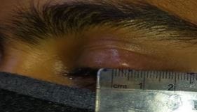
Figure 1: Circular firm swelling of size 0.5*0.5cm.

opthal_224_02.JPG
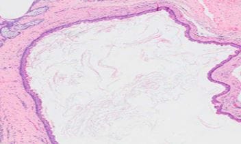
Figure 2: Histology - Trichilemmal cyst.

conjunctiva, caruncle, and pulp of the index finger. [8,9,10]. However, they are extremely rare on eyelids. However histologically trichilemmal cysts lack a granular cell layer.[11]. Approximately, 20% of the epithelial cysts are trichilemmal cysts and the other 80% are epidermoid.[12]. Very rarely, trichilemmal cysts can undergo malignant transformation. [5,6]. There are several types of benign and malignant lesions of the eyelids.


The common benign lesions are chalazion, epidermal inclusion cysts, seborrheic keratosis, and apocrine hidrocystomas. [11]. Both trichilemmal and epidermal cysts are keratinous cysts and usually have similar clinical presentations [13]. However, these cysts differ significantly in their manner of keratinization on histopathology. Trichilemmal cyst shows trichilemmal or abrupt keratinization without keratohyalin granules. The peripheral layers demonstrate a palisading arrangement, whereas cells close to the cyst cavity are swollen and filled with pale cytoplasm. The cyst cavity contains amorphous eosinophilic keratin. Foci of calcifications within the keratin occur in approximately 25% of cases [14]. Therefore, histopathological examination is important to differentiate between the two conditions. Approximately, 20% of the epithelial cysts are trichilemmal cysts and the other 80% are epidermoid.[12].

Conclusion

We hereby conclude that a trichilemmal cyst is extremely rare on the upper eyelid. And it may also occur in males; however, it is being reported more in females. The main modality of treatment remains excision however it is known to recur even after that so new modalities should be looked at. The trichilemmal cyst has to be differentiated from other benign lesions of the eyelid especially chalazion as it may turn malignant.

Reference

01. Adachi N, Yamashita T. Trichilemmal cyst of the scalp: MR imaging. AJR Am J Roentgenol. 1990 Feb;154(2):423-4. doi: 10.2214/ajr.154.2.2105043 [Crossref][PubMed][Google Scholar]

02. Ikizoglu, Guliz, et al. The use of the freer dissector for the removal of trichilemmal cysts. " International journal of dermatology 42. 5 (2003): 405-407. [Crossref][PubMed][Google Scholar]

03. Karuppannon, Bashkaran, et al. Trichilemmal cyst in the eyelid. Int J Ophthalmol 9. 8 (2009). [Crossref][PubMed][Google Scholar]

04. Meena, Manju, Ruchi Mittal, and Debarati Saha. Trichilemmal cyst of the eyelid: masquerading as recurrent chalazion. Case Reports in Ophthalmological Medicine 2012 (2012). [Crossref][PubMed][Google Scholar]

05. Rutty, G. N. , P. I. Richman, and J. H. E. Laing. Malignant change in trichilemmal cysts: a study of cell proliferation and DNA content. Histopathology 21.5 (1992): 465-468 [Crossref][PubMed][Google Scholar]

06. Lai, Tze Foon, et al. Trichilemmal carcinoma of the upper eyelid. Acta Ophthalmologica Scandinavica 81. 5 (2003): 536-538. [Crossref][PubMed][Google Scholar]

07. Kadri, Rajani, et al. Trichilemmal cyst of the bulbar conjunctiva: a rare presentation. Middle East African Journal of Ophthalmology 20. 4 (2013): 366. [Crossref][PubMed][Google Scholar]

08. Jakobiec, Frederick A. , Manisha Mehta, and Francis Sutula. Keratinous cyst of the palpebral conjunctiva. " Ophthalmic Plastic & Reconstructive Surgery 25. 4 (2009): 337-339 [Crossref][PubMed][Google Scholar]

09. Jakobiec, Frederick A. , et al. The white caruncle: sign of a keratinous cyst arising from a sebaceous gland duct. " Cornea 29. 4 (2010): 453-455 [Crossref][PubMed][Google Scholar]

10. Ikegami, Takeshi, et al. Trichilemmal cyst in the pulp of the index finger: a case report. Hand Surgery 8. 02 (2003): 253-255. [Crossref][PubMed][Google Scholar]

11. Bernardini, Francesco P. Management of malignant and benign eyelid lesions. Current opinion in ophthalmology 17. 5 (2006): 480-484. [Crossref][PubMed][Google Scholar]

12. Satyaprakash, Anita K. , Daniel J. Sheehan, and Omar P. Sangueeza. Proliferating trichilemmal tumors: a review of the literature." Dermatologic Surgery 33.9 (2007): 1102-1108 [Crossref][PubMed][Google Scholar]

13. Elenitsas, R. , and A. Halpern. Biopsy Techniques in Lever‘s Histopathology of the Skin, eight edition. " (1997): 3-50 [Crossref][PubMed][Google Scholar]

14. Leppard BJ, Sanderson KV. The natural history of trichilemmal cysts. Br J Dermatol. 1976 Apr;94(4):379-90. doi: 10.1111/j.1365-2133.1976.tb06115.x [Crossref][PubMed][Google Scholar]