E-ISSN:2456-6454
P-ISSN:2581-4907
RNI:MPENG/2017/74152

Case Report

CCRAVO

Tropical Journal of Ophthalmology and Otolaryngology

2021 Volume 06 Number 06 November December
Publisherwww.medresearch.in

Bilateral Combined central retinal artery and vein occlusion (CCRAVO) in COVID-19 patient

Ganekal S.1*, Dorairaj S.2, Ganekal V.3
DOI: https://doi.org/10.17511/jooo.2021.i06.04

1* Sunil Ganekal, Research Collaborator, Department of Ophthalmology, Mayo Clinic College of Medicine, , Florida, USA.

2 Syril Dorairaj, Fellowship, Department of Ophthalmology, Mayo Clinic College of Medicine, , Florida, USA.

3 Varun Ganekal, Research Assistant, Department of Ophthalmology, , Davangere, Karnataka, India.

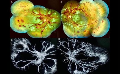
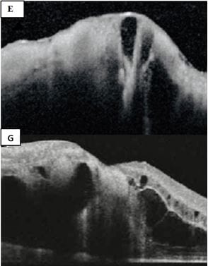
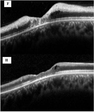
We report a case of a male patient who gave a history of fever, dry cough, dyspnea and he was tested positive for COVID-19 by Reverse Transcriptase-PCR from the nasopharynx. Presented with the sudden bilateral onset of decreased vision due to combined central retinal artery and vein occlusion (CCRAVO), the patient was treated with systemic medication including anticoagulant treatment and Intravitreal bevacizumab, and visual recovery was limited despite treatment.

Keywords: Combined central retinal artery and vein occlusion, COVID-19

Corresponding Author How to Cite this Article To Browse
Sunil Ganekal, Research Collaborator, Department of Ophthalmology, Mayo Clinic College of Medicine, , Florida, USA.
Email:
Sunil Ganekal, Syril Dorairaj, Varun Ganekal, Bilateral Combined central retinal artery and vein occlusion (CCRAVO) in COVID-19 patient. Trop J Ophthalmol Otolaryngol. 2021;06(06):128-131.
Available From
https://opthalmology.medresearch.in/index.php/jooo/article/view/222

Manuscript Received Review Round 1 Review Round 2 Review Round 3 Accepted
2021-12-06 2021-12-10 2021-12-17 2021-12-24 2021-12-31
Conflict of Interest Funding Ethical Approval Plagiarism X-checker Note
Nil Nil Yes 17%

© 2021by Sunil Ganekal, Syril Dorairaj, Varun Ganekaland Published by Siddharth Health Research and Social Welfare Society. This is an Open Access article licensed under a Creative Commons Attribution 4.0 International License https://creativecommons.org/licenses/by/4.0/ unported [CC BY 4.0].

Introduction

The incidence of thrombotic complications in patients with COVID-19 infection is elevated due to multiple factors and is associated with poorer outcomes. [1,2].  Hypercoagulability associated with COVID-19 has been described as a “sepsis-induced coagulopathy”. [3]. Such thrombotic events lead to retinal vascular occlusion. We report a rare case of combined retinal artery and vein occlusion in a COVID 19 patient, which has not been reported before.

Case Description

A 36-year-old man without any preexisting systemic comorbidities presented with fever, cough, and progressive dyspnea (SpO2-88%). RNA Reverse Transcriptase-PCR from the nasopharynx was positive for COVID-19. After two days of initial symptoms, the patient developed severe breathlessness, and the CT Scan showed patchy bilateral middle and lower field pulmonary infiltrates. He was shifted to ICU for acute hypoxemic respiratory distress and received COVID-19-directed therapy, including azithromycin, tocilizumab remdesivir and pulse steroid therapy. He remained in the intensive care for the entire seven days, and on stabilization, he was shifted from ICU to the medical ward for further management. On the thirteenth day since the onset of COVID-19 symptoms patient complained of sudden onset of painless profound loss of vision in both eyes. On examination, both eyes had visual acuity of light perception; the pupillary reaction was ill sustained in both eyes. Anterior segment examination and ocular motility were normal. Dilated fundus examination of both eyes showed mild disc edema, attenuated arteries and dilated veins with scattered peripapillary hemorrhages, cotton wool spots and retinal hemorrhages in all the quadrants. Right eye and Left eye fundus images showed disc edema, retinal hemorrhages, attenuated arteries, dilated veins, retinal edema with whitening. Area of retinal edema with whitening was noted more over the posterior pole, with this diagnosis of combined retinal artery and vein occlusion was made (Figure A & B). For further confirmation of diagnosis and management patient underwent OCT and fundus fluorescein angiography (FFA). FFA showed (Figure C& D) an absence of flow in both retinal artery and retinal

veins with an associated large area of capillary non-perfusion over the posterior pole and retinal periphery. Vessel wall staining was noted in a few quadrants.

opthal_222_01.JPG

OCT examination (Figure E& G) showed increased reflectivity in the inner retinal layers and diffuse macular edema (CMT RE-985µ and LE-916 µ). Choroidal details could not be appreciated.

opthal_222_02.JPG

The patient underwent workup for the hypercoagulable state (hereditary or acquired thrombophilia factors), vasculitic syndromes, blood hyperviscosity, and other recognized systemic vascular inflammatory disorders. Laboratory workup after the onset of visual symptoms revealed no pathological findings in blood analysis comprising  Blood sugar, lipid profile,


homocysteinemia, anti-cardiolipinIgM and IgG antibodies, and screening for genetic thrombophilia (Factor V Leiden and prothrombin mutations, antithrombin III and proteins C and S deficiencies), ANA, C-ANCA, P-ANCA, ds DNA, but showed elevated Inflammatory markers; D-Dimer (>1050 ng/ml; normal range <255ng/ml), serum ferritin(615.30 ng/ml; normal range 22-274ng/ml), CRP(13mg/L; Normal range<10mg/L), pro-calcitonin (0.16 ng/ml; normal ≤ 0.07 ng/ml). Coagulation profile showed; fibrinogen (546 mg/dl; normal range 200–400 mg/dl), prothrombin time (17.6 s; normal range 11.9–14.4 s), international normalized ratio (1.5; reference range 0.8–1.3), partial thromboplastin time (36 s; normal range 22–37 s). Haemogram showed mild lymphopenia (0.66 × 1000 µl/) and thrombocytosis (412 × 1000 µl/)).MRI of the brain and orbits and CT angiogram head and neck were normal. Transthoracic echocardiogram was unremarkable. Considering thrombotic microangiopathy as the cause for combined retinal vascular occlusion, the patient was started on anticoagulant enoxaparin 1 mg/kg twice daily therapy along with standard COVID-19 treatment protocol. The patient was treated with Intravitreal anti-VEGF (Bevacizumab, 1.25 mg/0.05 ml). Then a second intravitreal dose was given after four weeks and was asked to continue systemic medications, including tapering oral steroids as advised by the physician. The patient was followed up at one week, one month and two months. There was a partial improvement in best-corrected visual acuity in both eyes (1/60 both eyes) at the end of 2 months. At last, follow up of 2 months, OCT of both eyes showed resolution of macular edema but associated retinal thinning (Figure F & H).

opthal_222_03.JPG

Discussion

There are no published reports of combined bilateral retinal artery and vein occlusion in COVID- 19 patients, and these patients are at risk of presenting venous and arterial thrombotic events. COVID 19 infection is associated with coagulation activation and a disproportionate systemic inflammatory response. [4,5]. Whether the coagulation cascade is directly activated by the virus or the result of local or systemic inflammation is not entirely understood. The three main factors involved in the pathogenesis of coagulopathy in patients with COVID-19 are 1) Endotheliitis, which causes vasoconstriction, 2) hyper-inflammation, and cytokine storm, which activates clotting factors, 3) stasis and hypoxia activate coagulation mechanisms. [6] Ocular manifestations such as anterior uveitis, retinitis, and optic neuritis have been only documented in animal models.[7] Previous studies have reported CRAO, papillophlebitis and isolated CRVO secondary to COVID-19. [3,8].  In COVID-19, due to venous or arterial thromboembolic complications, evaluation for hypercoagulable disorders must be considered. In our patient, an interdisciplinary exploration process and a complete thrombophilia study were Performed, and the only findings were consistent with a hypercoagulable state induced by the COVID-19 infection and treated accordingly with anticoagulant (enoxaparin) and intravitreal bevacizumab but with limited post-treatment visual recovery.

Conclusion

Ophthalmologists must be prepared to treat vision-threatening retinal vascular occlusions in the intermediate stages of COVID-19. Evidence of venous and arterial thromboembolic events in these patients suggest that pharmacologic anticoagulation prophylaxis may benefit hospitalized patients with confirmed or highly suspected COVID-19.

Reference

01. Tang N, Li D, Wang X, Sun Z. Abnormal coagulation parameters are associated with poor prognosis in patients with novel coronavirus pneumonia. J Thromb Haemost. 2020 Apr;18(4):844-847. doi: 10.1111/jth.14768 [Crossref][PubMed][Google Scholar]


02. Llitjos JF, Leclerc M, Chochois C, Monsallier JM, Ramakers M, Auvray M, et al. High incidence of venous thromboembolic events in anticoagulated severe COVID-19 patients. J Thromb Haemost. 2020 Jul;18(7):1743-1746. doi: 10.1111/jth.14869 [Crossref][PubMed][Google Scholar]

03. Acharya S, Diamond M, Anwar S, Glaser A, Tyagi P. Unique case of central retinal artery occlusion secondary to COVID-19 disease. IDCases. 2020;21:e00867. doi: 10.1016/j.idcr.2020.e00867 [Crossref][PubMed][Google Scholar]

04. Wu Z, McGoogan JM. Characteristics of and Important Lessons From the Coronavirus Disease 2019 (COVID-19) Outbreak in China: Summary of a Report of 72 314 Cases From the Chinese Center for Disease Control and Prevention. JAMA. 2020 Apr 7;323(13):1239-1242. doi: 10.1001/jama.2020.2648 [Crossref][PubMed][Google Scholar]

05. Marietta M, Ageno W, Artoni A, De Candia E, Gresele P, Marchetti M, et al. COVID-19 and haemostasis: a position paper from Italian Society on Thrombosis and Haemostasis (SISET). Blood Transfus. 2020 May;18(3):167-169. doi: 10.2450/2020.0083-20 [Crossref][PubMed][Google Scholar]