E-ISSN:2456-6454
P-ISSN:2581-4907
RNI:MPENG/2017/74152

Research Article

Proptosis

Tropical Journal of Ophthalmology and Otolaryngology

2021 Volume 6 Number 6 November December
Publisherwww.medresearch.in

Clinical Profile of Proptosis in a Tertiary Eye Care Hospital in Central India

Sinha A.1*
DOI: https://doi.org/10.17511/jooo.2021.i06.01

1* Abha Sinha, Associate Professor, Department of Ophthalmology, BRLSABVM Medical College, Rajnandgaon, Chhattisgarh, India.

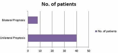
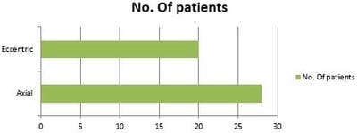
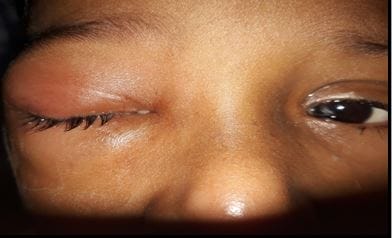
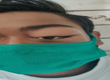
Introduction: Proptosis is the protrusion or forward displacement of one or both eyeballs from posterior pressure on the globe due to increased orbital volume either from intraorbital or intracranial space-occupying lesion. This study aims to find out the aetiology, clinical features, and its management. Material and Methods: Study setting- Department of ophthalmology, BRLSABVM Medical College, Rajnandgaon (C.G.), India. Study duration- July 2017 to June 2020. Type of study- Prospective study. Sampling methods- Universal sampling method. Sample size- 875 patients presenting with provisional diagnosis of proptosis over three years were included. Results: In the present study, 875 patients presented with a provisional diagnosis of proptosis; out of them in only 48 patients confirmed proptosis was seen. In the present study, it was found more common in males than females. The most common aetiology of proptosis in the present study was orbital cellulitis; among them, two had bilateral proptosis. The neoplasm was the second most common cause of proptosis, affecting 13 eyes of 11 patients. The most common symptoms were protrusion of the eye followed by eye pain & headache. Other symptoms were diminished vision, diplopia, diminished motility, epiphora, chemosis & proptosis. Conclusion: Present study showed that the most common cause of proptosis is orbital cellulitis. The etiology and pathogenesis of proptosis are diverse, which can be vision or life-threatening, highlighting the need for early and timely diagnosis of these cases, mandating various spectrum of investigations.

Keywords: Proptosis, Exophthalmometer, Orbital cellulitis

Corresponding Author How to Cite this Article To Browse
Abha Sinha, Associate Professor, Department of Ophthalmology, BRLSABVM Medical College, Rajnandgaon, Chhattisgarh, India.
Email:
Abha Sinha, Clinical Profile of Proptosis in a Tertiary Eye Care Hospital in Central India. Trop J Ophthalmol Otolaryngol. 2021;6(6):108-113.
Available From
https://opthalmology.medresearch.in/index.php/jooo/article/view/219

Manuscript Received Review Round 1 Review Round 2 Review Round 3 Accepted
2021-10-27 2021-10-29 2021-11-05 2021-11-12 2021-11-19
Conflict of Interest Funding Ethical Approval Plagiarism X-checker Note
Nil Nil Yes 18%

© 2021by Abha Sinhaand Published by Siddharth Health Research and Social Welfare Society. This is an Open Access article licensed under a Creative Commons Attribution 4.0 International License https://creativecommons.org/licenses/by/4.0/ unported [CC BY 4.0].

Introduction

Proptosis is the protrusion or forward displacement of one or both eyeballs from posterior pressure on the globe due to increased orbital volume either from intraorbital or intracranial space-occupying lesion [1]. It may be caused by the local orbital problem to infiltrative disease and spread from contiguous sites, including the nasopharynx, paranasal sinuses and sometimes distant structures. The etiological spectrum of proptosis ranges from inflammation, infection, neoplasia to endocrine and vascular disorders [2]. The etiology and treatment of proptosis are variable. There is significantly less study on the various aspects of proptosis in this part of central India. This study aims to find out the aetiology, clinical features, and its management. When aetiologies are known, it will help in early suspicion, diagnosis and its treatment. Recognizing the various causes of proptosis is important as many can be vision-threatening or life-threatening.

Material and Methods

Study setting- Department of ophthalmology, BRLSABVM Medical College, Rajnandgaon (C.G.), India.

Study duration- July 2017 to June 2020

Type of study- Prospective study.

Sampling methods- Universal sampling method.

Sample size- 875 patients presenting with with provisional diagnosis of proptosis over three years were included.

Inclusion criteria- Patients that presented with proptosis at the eye O.P.D., those admitted to the inpatient department of ophthalmology and referred from other departments.

Exclusion criteria- Those who are not willing to participate in the study.

Data collection & Surgical procedure- Proptosis was defined by exophthalmometry (Luedde’s exophthalmometer) with a value of >21 mm or a difference of >2 mm between both eyes. The gender and age distribution of cases were recorded. A detailed history and clinical examination of all patients were made. Measurement of proptosis was done with the help of an exophthalmometer. Relevant necessary laboratory investigations, orbital

imaging by either C.T. or M.R.I. was done in all patients. Patients were treated either medically or surgically according to the standard protocol.

History of pain was taken, if present at rest or with eye movement. Distribution of pain, whether it is constant or intermittent, was asked. History about the progression of proptosis was enquired.

In clinical examination, all patients examined were evaluated for the displacement of globe superiorly, inferiorly, medially, laterally or axially. An exophthalmometer was used to quantify the distance from the orbital rim to the corneal surface of each eye to assess the symmetry of globe protrusion. Proptosis was examined for any pulsation or bruit. Any changes in the colour of the skin of the eyelids, swelling of the eyelids were noted. Patients were asked to show the last photographs. To assess the optic nerve function, the patient's best-corrected visual acuity was recorded. Pupils were examined for a relative afferent pupillary defect, and colour vision was evaluated using a colour vision chart. Fundus examination was done to see for papilloedema or optic disc pallor. Relevant laboratory investigations like C.B.C., PT/PTT and thyroid profile, Orbital ultrasonography, orbital imaging by either C.T. &/or M.R.I. was done in all patients to confirm the diagnosis & to ascertain the aetiology for proptosis. The case details were recorded & classified based on etiology as inflammatory, vascular, traumatic, endocrine & neoplastic.

Ethical consideration & permission- Ethical considerations were met through the institutional ethical committee. Informed consent was obtained from all the participants.

Statistical Analysis- Data was compiled in M.S. excel and checked for its completeness and correctness. Then it was analyzed by using a suitable statistical software package.

Results

In the present study, 875 patients presented with a provisional diagnosis of proptosis. Out of them in only 48 patients confirmed proptosis was seen. Patients presenting with proptosis belonged to different age groups. The present study was found more common in males (31 patients) than females (17). [Table 1].


Proptosis was unilateral in 40 patients & bilateral in 8 patients (Fig-1).

The most common etiology of proptosis in the present study was orbital cellulitis (14 cases); among them, two had bilateral proptosis. The neoplasm was the second most common cause of proptosis, affecting 13 eyes of 11 patients. 2 patients had bilateral proptosis. Thyroid ophthalmopathy was found in 8eyes of 5 patients, of which three patients had bilateral proptosis. In 3 patients, the cause of proptosis was pseudotumour orbit. Other causes of proptosis were mucormycosis in 1 patient, vascular lesions in 3 patients & mucocele in 2 patients. Trauma was responsible for 2 cases of unilateral proptosis. In 4 patients etiology of unilateral proptosis was congenital lesions. In 1 patient, sarcoidosis was the cause of bilateral proptosis. The diagnosis was not known in 2 patients with unilateral proptosis. [Table-2] 

It was axial in 28 cases & eccentric (nonaxial) in 20 cases [Table-3, Figure-2].

The most common symptoms were protrusion of the eye (48 cases) followed by eye pain (34 cases) & headache (30 cases). Other symptoms are diminished vision, diplopia, diminished motility, epiphora, chemosis & proptosis. [Table-4]

Patients with orbital cellulitis presented with proptosis, restricted extraocular motility, conjunctival congestion. In some patients with orbital cellulitis, relative afferent pupillary defect, optic nerve, edema & pallor was seen. Patients of orbital cellulitis were managed with IV broad-spectrum antibiotics for 7-10 days, nonsteroidal anti-inflammatory drugs, topical antibiotics and topical lubricating drops. In all patients with orbital cellulitis, proptosis subsided, and in 12 patients, visual improvement occurred after treatment. In 2 patients, there was no visual improvement due to optic nerve involvement. The second most common cause of proptosis was the orbital tumour. Most were orbital malignancies viz, retinoblastoma (1 patient), squamous cell carcinoma (3 patients), lymphoma (1 patient), adenocarcinoma of the lacrimal gland (1 patient). Some were benign tumours, meningioma (1 patient), haemangioma (1patient), lymphangioma (1 patient), optic nerve gliomas, pleomorphic adenoma of lacrimal glands. These orbital tumours were diagnosed clinically and with orbital imaging like CT/MRI.

Table-1: Age & Gender wise distribution of patients presenting with proptosis

Age in years No. %
0-15 4 8
16-30 7 15
31-45 13 27
46-60 11 23
>60 13 27
Gender    
 Male 31 64.6
Female 17 35.4

Table-2: Etiology of proptosis

S.N. Etiology No. %
1 Orbital cellulitis 14 29.16
2 Neoplasm 11 23
3 Thyroid ophthalmopathy 5 10.42
4 Pseudotumour 3 6.25
5 Mucormycosis  1 2
6 Vascular lesions 3 6.25
7 Trauma(Orbital/Retrobulbar hematoma)  2 4.2
8 Mucocoele  2 4.2
9 Sarcoidosis 1 2
10 Congenital lesions  4 8.33
11 Undiagnosed cases  2 4.2

Table-3: Types of proptosis

S. N. Proptosis types No.
1. Axial 28
2. Eccentric 20

Table-4: Presenting Symptoms 

S.N. Symptoms No. (48)
1 Proptosis 48
2 Diminished vision 20
3 Diplopia 3
4 Diminished motility 6
5 Epiphora  2
6 Headache 30
7 Chemosis 25
8 Orbital mass 5
9 Eye pain 34
10 Ptosis 4

This helped in the localization of the lesions, and etiology could be suspected with the help of C.T. & M.R.I. In the present study, the thyroid ophthalmopathy was the etiology of proptosis in 8 eyes of 5 patients. The diagnosis of thyroid ophthalmopathy was based on clinical (eyelid retraction, proptosis) and radiological


signs like orbital fat expansion & fusiform rectus muscle enlargement with sparing of muscle tendons. Lab investigations like T3, free T4 & TSH levels were also done in all thyroid eye disease patients& for the control thyroid hormone levels, patients were referred to the medical department. For mild disease, lubricant eye drops were given for an ocular surface disease. In 3 patients, the cause of proptosis was orbital pseudotumor, where no known etiology was found.

In 3 patients,the cause of proptosis was orbital pseudotumor,where no known etiology was found. Patients presented with myositis, pain, periorbital edema, ptosis, chemosis, proptosis & decreased ocular motility. The diagnosis was made based on clinical findings, orbital imaging with enhancing lesions, rectus muscle enlargement with tendon involvement and scleral enhancement with prompt response to systemic steroid administration. In 2 patients, trauma was found responsible for the proptosis. Patients presented with pain, proptosis, diminished vision, reduced ocular motility, increased intraocular pressure and subconjunctival hemorrhage. Patients were managed with lateral canthotomy with inferior and superior cantholysis. Congenital lesions were seen in 4 patients, vascular lesions were found in 3 eyes of 3 patients. In 2 patients, mucocele and one, sarcoidosis was responsible for proptosis. In 2 cases of unilateral proptosis, the diagnosis was not confirmed. [Figure. 3& 4]

opthal_219_01.JPG

Fig-1: Unilateral (40) and Bilateral proptosis (8)

opthal_219_02.JPG

Fig-2: Eccentric (20) and Axial proptosis (28)

opthal_219_03.JPG

Fig-3: A case of orbital cellulitis

opthal_219_04.JPG

Fig-4: A case of pseudotumour orbit

Discussion

The bulging of the eyeball anteriorly from its bony cavity by a distance of about 14-20 mm due to various etiologies results in proptosis [3,5]. It results from many pathologies that can either produce threats to the vision or might be life-threatening, so its etiology needs to be carefully evaluated. In this study, the prevalence of proptosis was 5.5%. In the present study, proptosis was most prevalent in the age group of 31-45 yrs. This was similar to another Indian study by Sripurapu S, Raju TJ et al., where proptosis was more common in this age group [6]. In Majeko dunmi study, however, 87% of proptosis was found in patients 30 yrs of age and below, with 47% of proptosis occurring in children < 15 yrs [7]. This significant difference might be because her research was skewed towards children. Our study also showed a male preponderance. Similar findings were demonstrated by Khan et al. [13]. Loganathan and Radhakrishnan [8]. and Sharma et al. ([9]. Whereas Satpute and Chingsuin gamba looking at proptosis in general, found equal distribution among both


sexes [10]. Zaidi SH et al. showed a female preponderance [11]. Unilateral proptosis was more common in our study, as was found by S. Guthoupe J.D. and Hochman M [4]. The axial direction of proptosis was noticed maximally in this study which is similar to the observation done by Loganathan M, Radhakrishnan M et al. [8]. Orbital cellulitis was the most common cause of proptosis in our study; similar results were obtained in a study done by Majekodunmi S et al. in Nigeria  & a study done by Sultana A et al. in South India [7,12]. In a study done by Masud et al. in Peshawar, infection was the number three cause of proptosis (2). The orbital tumour is the second most common cause of proptosis in this study. Khan et al., in their research, found orbital tumours as the cause of proptosis in two-thirds of their patients [13]. also, Ogbeide and Theophilus in Benin city reported that tumour was the most common cause of proptosis. [14]. Thyroid ophthalmopathy was the third most common cause of proptosis found in our study, which showed the frequent occurrence of bilateral proptosis in thyroid ophthalmopathy, which is consistent with the findings by Sabharwal et al. [15]. In the present study, trauma due to a road traffic accident presenting as orbital hemorrhage was an important cause of proptosis which was similar to the results by Ogbeide and Theophilus in Benin city Nigeria (14). Other reasons for proptosis in our study included Vascular lesions, Congenital lesions, Mucormycosis, Sarcoidosis etc. In 2 cases, the diagnosis was not confirmed.

Conclusion

The present study showed that the most common cause of proptosis is orbital cellulitis. In other studies most common cause was neoplasm. The etiology and pathogenesis of proptosis are diverse, which can be vision or life-threatening, highlighting the need for early and timely diagnosis of these cases, mandating various spectrum of investigations. C.T. Scan was a vital imaging tool for evaluating a claim of proptosis, but histopathological examination gives a definitive diagnosis of the exact aetiology.

Acknowledgements: The authors would like to thank the staff and faculty of the Department of Ophthalmology, BRLSABVMMC, Rajnandgaon (C.G.), India, for their support during the entire study period without which this study wouldn’t have been possible.

Reference

01. Kamminga N, Jansonius NM, Pott JW, Links TP. Unilateral proptosis: the role of medical history. Br J Ophthalmol. 2003 Mar;87(3):370-1. doi: 10.1136/bjo.87.3.370 [Crossref][PubMed][Google Scholar]

02. Masud MZ, Babar TF, Iqbal A, Khan MT, Zaffar ul Islam, Khan MD. Proptosis: etiology and demographic patterns. J Coll Physicians Surg Pak. 2006 Jan;16(1):38-41. [Crossref][PubMed][Google Scholar]

03. Calcaterra TC, Trapp TK. Unilateral proptosis. Otolaryngol Clin North Am. 1988 Feb;21(1):53-63. [Crossref][PubMed][Google Scholar]

04. Khan, N. H. , et al. Unilateral proptosis: a local experience. Biomedica 20.2 (2004): 1114-6 [Crossref][PubMed][Google Scholar]

05. Ali, Zulfiqar, and Anjum Khawar. Non-thyroidal Unilateral Proptosis. The Journal of Surgery PIMS 5 (1993): 39-40. [Crossref][PubMed][Google Scholar]

06. Siripurapu, Suman, T. Jaya Raju, and K. Chandra Sekhar. "A Study on Clinical Correlation of Orbital Diseases Interventional and non-Interventional Diagnostic Procedures. " Indian Journal of Public Health Research & Development 7.1 (2016) [Crossref][PubMed][Google Scholar]

07. Majekodunmi S. Unilateral proptosis in Nigerians: causes and differential diagnosis. Can J Ophthalmol. 1982 Oct;17(5):203-6. [Crossref][PubMed][Google Scholar]

08. Loganathan, M. , and M. Radhakrishnan. An etiological analysis of childhood proptosis. Journal of Evolution of Medical and Dental Sciences 3.22 (2014): 6158-6163 [Crossref][PubMed][Google Scholar]

09. Sharma P, Tiwari PK, Ghimire P. G. , Ghimire P. Role of computed tomography in evaluation of proptosis. " Nepal Journal of Medical Sciences 2.1 (2013): 34-37 [Crossref][PubMed][Google Scholar]

10. Satpute, Kishor K. , and Y. Chingsuingamba. A retrospective analysis of presentation and management outcome of proptosis in a tertiary care centre of North-East, India-a case series. J Dent Med Sci 5 (2013): 30-2 [Crossref][PubMed][Google Scholar]


11. Zaidi SH. Unilateral proptosis in E. N. T. practice. J Pak Med Assoc. 1991 Oct;41(10):248-50 [Crossref][PubMed][Google Scholar]

12. Sultana, Aliya. Analysis of prevalence and etiology of proptosis at a tertiary care centre of South, India. Ann Int Med Dent Res 3 (2017): 5-8. [Crossref][PubMed][Google Scholar]

13. Khan NH, Moin M, Khan MA, Azhar Hameed. Unilateral proptosis: a local experience. Biomedica 20. 2 (2004): 1114-6. [Crossref][PubMed][Google Scholar]

14. Ogbeide, Ehimwenma, and Akhigbe Oriazo Theophilus. "Computed tomographic evaluation of proptosis in a Southern Nigerian tertiary hospital. " Sahel Medical Journal 18. 2 (2015): 66. [Crossref][PubMed][Google Scholar]

15. Sabharwal, K. , A. L. Chouhan, and S. Jain. C.T. evaluation of proptosis. Indian Journal of Radiology and Imaging 16.4 (2006) [Crossref][PubMed][Google Scholar]