E-ISSN:2456-6454
P-ISSN:2581-4907
RNI:MPENG/2017/74152

Research Article

Eye Multifactorial Disease

Tropical Journal of Ophthalmology and Otolaryngology

2021 Volume 6 Number 5 September October
Publisherwww.medresearch.in

Clinical Profile of Dry Eye in a Tertiary Care Hospital of Central India

Sinha A.1*
DOI: https://doi.org/10.17511/jooo.2021.i05.02

1* Abha Sinha, Associate Professor, Department of Ophthalmology, BRLSABVM GMC, Rajnandgaon, Chhattisgarh, India.

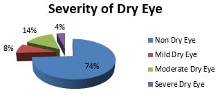
Introduction: Dry eye is a multifactorial disease of the tear film and ocular surface that results in symptoms of discomfort, visual disturbance and tears film instability with potential damage to the ocular surface. Dry eye disease is a frequent cause of ocular irritation that leads the patients to seek ophthalmic care. Material and Methods: This study was conducted in the department of ophthalmology, BRLSABVM Medical College, Rajnandgaon (C.G.), India, from Oct 2018 to Sep 2020. One thousand six hundred twenty-three patients presenting with ocular surface symptoms over two years were included in the study. Patients above the age of 20 years having symptoms of irritation, tearing, burning, stinging, Foreign body sensation, mild itching, photophobia, blurry vision, redness, increased frequency of blinking for the one-month duration were included. Data was compiled in M.S. excel & analyzed by using a suitable statistical software package. Results: In this study, 1623 patients presenting with ocular surface symptoms were taken. The prevalence of dry eye disease in our study is 26% (422). Dry eye was most prevalent among 20-40 yrs of age. The majority of dry eye was more in the male gender than female. Occupation having regular computer use was more predisposed to develop dry eye disease. In the present study, the most common ocular morbidity associated with dry eye disease is Meibominitis. Conclusion: Increasing prevalence of dry eye cases in the younger age group is attributed to the increased use of computer & other visual display terminals like laptops, smartphones & tablets etc., by these age groups.

Keywords: Dry eye, Visual acuity, Visual discomfort, Tears film

Corresponding Author How to Cite this Article To Browse
Abha Sinha, Associate Professor, Department of Ophthalmology, BRLSABVM GMC, Rajnandgaon, Chhattisgarh, India.
Email:
Abha Sinha, Clinical Profile of Dry Eye in a Tertiary Care Hospital of Central India. Trop J Ophthalmol Otolaryngol. 2021;6(5):93-98.
Available From
https://opthalmology.medresearch.in/index.php/jooo/article/view/215

Manuscript Received Review Round 1 Review Round 2 Review Round 3 Accepted
2021-09-07 2021-09-09 2021-09-16 2021-09-23 2021-09-30
Conflict of Interest Funding Ethical Approval Plagiarism X-checker Note
Nil Nil Yes 16%

© 2021by Abha Sinhaand Published by Siddharth Health Research and Social Welfare Society. This is an Open Access article licensed under a Creative Commons Attribution 4.0 International License https://creativecommons.org/licenses/by/4.0/ unported [CC BY 4.0].

Introduction

Dry eye disease is a prevalent condition in recent years that causes a varying degree of ocular discomfort and disability. It is a multifactorial disease of the tear film and ocular surface that results in symptoms of pain, visual disturbance and tears film instability with potential damage to the ocular surface. It is accompanied by the increased osmolality of the tear film and inflammation of the ocular surface [1]. Dry eye disease is differentiated from other ocular diseases and subcategorized into aqueous deficient and evaporative dry eye disease [2]. Aqueous deficient dry eye disease results from the reduced lachrymal secretion in the presence of a normal rate of tear evaporation, while evaporative dry eye disease results from excessive waterlessness through evaporation from the tear film in the presence of normal lachrymal function [3]. Dry eye disease is a frequent cause of ocular irritation that leads the patients to seek ophthalmic care. It presents with a wide variety of nonspecific symptoms hence often unrecognized causing great frustration to the pts and ophthalmologists.

In many cases, dry eye disease leads to significant visual morbidity. Staring at a computer monitor or digital displays for hours has become a part of the modern workday, and inevitably all of that staring can put a real strain on our eyes [4]. Many such individuals then experience dry eye symptoms constantly and severely affect their quality of life [5].

Aim of Study

To estimate the proportion of dry eye diseases and their clinical profile in patients presenting with ocular surface symptoms to ophthalmology O.P.D. in a tertiary care Centre over 2 Years.

Material and Methods

Study setting- Department of ophthalmology, BRLSABVM Medical College, Rajnandgaon (C.G.), India

Study duration- October 2018 to September 2020

Type of study- Prospective study

Sampling methods- Universal sampling method

Sample size- 1623 patients presenting with ocular surface symptoms over two years were included.

Inclusion criteria- Patients above the age of 20 years having symptoms of irritation, tearing, burning, stinging, Foreign body sensation, mild itching, photophobia, blurry vision, redness, increased frequency of blinking for the one-month duration were included.

Exclusion criteria- Patients who had suffered from corneal or conjunctival infections, contact lens users and those who had undergone extraocular or intraocular surgery were excluded from the study.

Data collection & Surgical procedure- A history of all pts were taken in detail, which includes Age, Sex, Occupation History of allergic eye diseases, Topical & Systemic Medications usage, Ocular Surgical History, Ocular Surface Disease, Chemical Injury, systemic inflammatory disease like Sjogren Syndrome, RheumatoidArthritis & any Chronic viral infections. In addition, history of visual display terminal usage, including computers, tablets, laptops & smartphones, was also elicited. Screening of dry eye diseases was done using the O.S.D.I. questionnaire. O.S.D.I (ocular surface disease index) questionnaire contains 3sections. Section 1 is based on the relative frequency of occurrence of each symptom(gritty feeling in the eye, light sensitivity and blurred vision). Section 2 includes questions indicating limitations on certain activities(reading, driving at night, watching television). Section 3 is based on environmental conditions(wind, low humidity and air conditioning) on eyes. The grading of dry eye was made based on O.S.D.I.score as mild 13-22, moderate 23-32 & severe 33-100. Visual acuity of each patient was taken using Snellen's Chart. A thorough slit lamp examination of the anterior segment was done.

Eyelids are examined for trichiasis, entropion &ectropion. Eyelid margins were examined for abnormalities of meibomian glands and character of meibomian gland secretions (e.g. turbid, thickened, foamy). Puncta were examined for their position &patency. Tear film was examined for the height of the meniscus, any debris & mucous strands. Inferior fornix and tarsal conjunctiva were examined for mucous threads, scar,follicles&symblepharon formation. Bulbar conjunctiva was examined for Bitots spot, keratinization &papillae. Cornea was examined for punctuate epithelial erosions & filamentary keratopathy etc. Tear film breaks uptime, and ocular surface staining with fluorescein was done first. The Schirmer test followed this.


Tear film break up time [1, 2]

To test for the tear film break up time, a 2% fluorescein strip was instilled into the conjunctival sac. Patients were instructed to blink naturally three times and to cease blinking until required. The time-lapse between the last blink and the appearance of the first randomly distributed dark spots on the cornea was noted under the cobalt blue filter of the slit lamp. A value <10 seconds was taken as abnormal.

Staining pattern with fluorescein dye of conjunctiva and cornea was noted and graded as Nil, Mild or diffuse.

Lissamine Green Staining [1, 2]

The impregnated strip was moistened with tear substitute and applied to the lower palpebral conjunctiva. After 15 s of staining, conjunctiva and cornea were examined under a red-free filter of the slit lamp. The ocular surface was divided into three zones: Nasal bulbar conjunctiva, temporal bulbar conjunctiva and cornea were evaluated and graded from 0 to 3. In mucin deficiency dry eye, a score was found more than 3.

Schirmer's test[1, 2]

Thirty minutes after the staining, Schirmer's test was performed. Proparacaine was applied to both eyes. A narrow filter paper strip (Whatman Filter paper No.41) was used in the lower fornix at the junction of lateral 1/3rd and medial 2/3rd. Pt was allowed to blink. The schemer's strip was removed after 5 minutes. Amount of wetting was noted. Value <5mm. of the strip, wetting was taken as abnormal. Blood investigations like R.A. factor, ANA& Thyroid function tests were done.

Ethical consideration & permission- Ethical considerations were met through the institutional ethical committee. Informed consent was obtained from all the participants.

opthal_215_01.JPG

Fig. 1 Tear Film Break Up Time

opthal_215_02.JPG

Fig 2 Schirmer Test [1, 2]

Statistical Analysis- Data was compiled in M.S. excel and checked for its completeness and correctness. Then it was analyzed by using a suitable statistical software package.

Results

In this study, 1623 patients presenting with ocular surface symptoms were taken who were administered the O.S.D.I questionnaire. The prevalence of dry eye disease in our study is 26% (422). [Table-1]

Table 1-Demographic Profile of patients (1623) who were administered the O.S.D.I Questionnaire 

 Demographic      Characteristics No. %
Age(Yrs)
20-40 826 51%
40-60 548 34%
>60 249 15%
Sex
Male 975 60%
Female 649 40%

Dry eye was most prevalent among 20-40 yrs of age with a mean age of 32 yrs. In the present study, the prevalence of dry eye was more in the male gender than female. Occupation having regular computer use was more predisposed to develop dry eye disease. The majority of dry eye is positively correlated to the hours of computer usage/day. The use of visual display terminals like mobile, tablets etc., has been associated with a decreased maximum blink interval which leads to dry eye symptoms. Systemic diseases associated with dry eye in the present study was Hypertension(  6.2%), diabetes Mellitus( 2.4% ), thyroid disease( 2.1%), Sjogren's syndrome(1.6%), Rheumatoid arthritis(1.4%), Stevens Johnson Syndrome(0.4%)  & other connective tissue disorders(2%) Among them


Sjogren's syndrome, Stevens-Johnson Syndrome patients presented with severe dry eye in our study. [Table-2]

Table-2 - The demographic details of patients with dry eye diseases (422 patients) 

Demographic Characteristics No. %
Age(Yrs)
20-40 213 50%
40-60 182 43%
>60 27 7%
 Sex
Male 318 75%
Female 104 25%
Occupation
Desk Job With Computer Use 182 43%
House Wife 86 21%
Agricultural Workers 45 10.5%
Labourer/Factory Workers 74 17.5%
Others 35 8%

Fig 3. The severity of Dry Eye

opthal_215_03.JPG

Figure 3 shows the severity of dry eye among pts included in the present study according to grade. Most of the pts had a moderate dry eye (14%). [Figure-3]

In the present study, the most common ocular morbidity associated with dry eye disease is Meibominitis. [Table-3]

Table-3. Ocular Morbidity in Dry Eye Patients 

Disorder No. %
Blepharitis 21 5%
Meibominitis 204 48%
Xerosis 38 9%
Pterygium 96 23%
Bitot Spots 7 1.7%
Corneal filament 13 3%
Corneal Opacity 21 5%
Symblepharon 4 1%
Ectropion 11 2.6%
Trichiasis 7 1.7%

In our study Tear Film break up time was positive, i.e. <10 sec. in 216 out of 240 patients (90%). Dry eye with lipid layer deficiency was found in 108 patients (45%). Mucin layer deficient dry eye was present in81 patients (34%). Aqueous layer deficient dry eye was in 51 patients (21%). [Table-4]

Table-4. Dry Eye Test Result 

Test Positive % Of Dry Eye
Tear Film break uptime 216 90%
Slit-lamp examination (Lipid layer deficiency) 108 45%
Lissamine green staining (Mucin layer deficiency) 81 34%
Schirmer's test(Aqueous layer deficiency) 51 21%

Discussion

The prevalence of dry eye in the present study was 26% (422 patients of a dry eye out of 1623patients) based on the O.S.D.I. questionnaire. Dry Eye Disease in India is higher than the global prevalence and ranges from 18.4% to 54.3 % [6, 7]. The vast disparity in the prevalence of dry eye disease may be due to two factors- First, the geographical location of the study population and secondly, there is no standardization of the selected population, dry eye questionnaires, objective tests and dry eye diagnostic criteria [8,9]. In our study, Dry Eye Disease is most prevalent among 20-40 years of age. Increased prevalence of Dry Eye Disease in these age groups might be attributed to the increasing trend of computers & other visual display terminals like smartphones, tablets etc. these. Blue light emitted from smartphones can damage the corneal epithelium. Sahai and Malik reported a similar trend but relative peak in 3rddecade (20%). Rege et al. study said similar trends but a relative peak in the 6th decade, while Gupta et al. study reported a rise in the 8th decade (41.2%) [6, 10, 11]. Among genders, males (75%) were affected more commonly than females(25%). Similarly Jeewan Singh Titiyal study showed a significantly higher occurrence of Dry Eye Disease in males that might be due to the neglected attitude of females regarding treatment for their health problems. WhereasGuptaetal study based on O.S.D.I. questionnaire reported a higher prevalence of dry eye in females (27.4%) than males (11.8%).Mossetal& Beijing eye study also reported a higher prevalence in females than males [6, 12,13, 14,15]. Occupation having regular use of computers is associated with the development of


Dry Eye Disease in 162cases with more severe Dry Eye Disease in 18 cases. This is due to low humidity in the indoor environment & Air-conditioned rooms, which lead to Dry Eye Disease. Computer use for more than 8hrs a day has been reported as a significant risk factor for Dry Eye Disease, mainly attributed to a decrease in blink rate while using these devices, thereby hampering the uniform distribution of the tear film over the ocular surface. [12]. Jeewan SinghTitiyaletal study reported a similar trend with a higher occurrence of Dry Eye Disease in computer users & having more severe Dry Eye Disease.  In our research, the most common symptom seen in pts with Dry Eye Disease was Itching followed by a burning sensation. Meibomianitis (48%) is the most common ocular morbidity found in dry eye patients. Tear film breakup time was found positive in 216 dry eye patients, an essential diagnostic tool for dry eye. The most common tear film abnormality in the present study is lipid layer deficiency (45%), followed by mucin (34%) and aqueous layer deficiency. (21%) Schirmer test, which indicates Aqueous tear deficiency was positive only in 51 (21%) of dry eye cases. Due to the poor correlation between patient symptoms & clinical signs of dry eye. Multiple tests are done to diagnose dry eye. Symptoms, Questionnaires and objective dry eye tests are used to establish a diagnosis of dry eye syndrome and assess the effects of treatments or grade disease severity as published in studies by Schein et al. [13].

Conclusion

The prevalence of Dry eye is 26% in the present study, which suggests that dry eye disease is an under-diagnosed disease. Only with the help of tear parameters diagnosis of dry eye can be misleading. So combined approach with questionnaires & various tear parameters can be used to diagnose dry eye cases. Increased use of visual display terminals like smartphones for long hours can disturb the tear film and damage the ocular surface causing many dry eye symptoms. So there should be cautious use of these devices amongst the younger age group.

What does this study add to existing knowledge?

The increasing prevalence of dry eye cases in the younger age group is attributed to the increased use of computers & another visual display terminal.

Acknowledgements

The authors would like to thank the staff and faculty of the Department of Ophthalmology, BRLSABVMMC, Rajnandgaon (C.G.), India, for their support during the entire study period without which this study wouldn't have been possible. 

Author Contributors details in the study

  Author 1
Concepts Yes
Design Yes
Literature Search Yes
Clinical Studies Yes
Experimental Studies, Data Acquisition Yes
Data Analysis Yes
Statistical Analysis Yes
Manuscript Preparation Yes
Manuscript Editing Yes
Manuscript Review Yes

Reference

01. The definition and classification of dry eye disease: report of the Definition and Classification Subcommittee of the International Dry Eye WorkShop (2007). Ocul Surf. 2007 Apr;5(2):75-92. doi: 10. 1016/s1542-0124(12)70081-2 [Crossref][PubMed][Google Scholar]

02. Craig JP, Nichols KK, Akpek EK, Caffery B, Dua HS, Joo CK, et al. TFOS DEWS II Definition and Classification Report. Ocul Surf. 2017 Jul;15(3):276-283. doi: 10.1016/j.jtos.2017.05.008 [Crossref][PubMed][Google Scholar]

03. Bron AJ, de Paiva CS, Chauhan SK, Bonini S, Gabison EE, Jain S, et al. TFOS DEWS II pathophysiology report. Ocul Surf. 2017 Jul;15(3):438-510. doi: 10.1016/j.jtos.2017.05.011 [Crossref][PubMed][Google Scholar]

04. Uchino M, Uchino Y, Dogru M, Kawashima M, Yokoi N, Komuro A, et al. Dry eye disease and work productivity loss in visual display users: the Osaka study. Am J Ophthalmol. 2014 Feb;157(2):294-300. doi: 10.1016/j.ajo.2013.10.014 [Crossref][PubMed][Google Scholar]

05. Miljanović B, Dana R, Sullivan DA, Schaumberg DA. Impact of dry eye syndrome on vision-related quality of life. Am J Ophthalmol. 2007 Mar;143(3):409-15. doi: 10.1016/j.ajo.2006.11.060 [Crossref][PubMed][Google Scholar]


06. Gupta N, Prasad I, Jain R, D'Souza P. Estimating the prevalence of dry eye among Indian patients attending a tertiary ophthalmology clinic. Ann Trop Med Parasitol. 2010 Apr;104(3):247-55. doi: 10.1179/136485910X12647085215859 [Crossref][PubMed][Google Scholar]

07. Shah S, Jani H. Prevalence and associated factors of dry eye: Our experience in patients above 40 years of age at a Tertiary Care Center. Oman J Ophthalmol. 2015 Sep-Dec;8(3):151-6. doi: 10.4103/0974-620X.169910 [Crossref][PubMed][Google Scholar]

08. 2007 Report of the International Dry eye Workshop(DEWS)ocul. surf 2007,5:65-199. . 2015 Sep-Dec;8(3):151-6. doi: 10.4103/0974-620X.169910 [Crossref][PubMed][Google Scholar] [Crossref][PubMed][Google Scholar]

09. Shimmura S, Shimazaki J, Tsubota K. Results of a population-based questionnaire on the symptoms and lifestyles associated with dry eye. Cornea. 1999 Jul;18(4):408-11. doi: 10.1097/00003226-199907000-00003 [Crossref][PubMed][Google Scholar]

10. Sahai A, Malik P. Dry eye: prevalence and attributable risk factors in a hospital-based population. Indian J Ophthalmol. 2005 Jun;53(2):87-91. doi: 10.4103/0301-4738.16170 [Crossref][PubMed][Google Scholar]

11. Rege A, Kulkarni V, Puthran N, Khandgave T. A Clinical Study of Subtype-based Prevalence of Dry Eye. J Clin Diagn Res. 2013 Oct;7(10):2207-10. doi: 10.7860/J.C.D.R./2013/6089.3472 [Crossref][PubMed][Google Scholar]

12. Uchino M, Yokoi N, Uchino Y, Dogru M, Kawashima M, Komuro A, et al. Prevalence of dry eye disease and its risk factors in visual display terminal users: the Osaka study. Am J Ophthalmol. 2013 Oct;156(4):759-66. doi: 10.1016/j.ajo.2013.05.040 [Crossref][PubMed][Google Scholar]

13. Schein OD, Tielsch JM, Munõz B, Bandeen-Roche K, West S. Relation between signs and symptoms of dry eye in the elderly. A population-based perspective. Ophthalmology. 1997 Sep;104(9):1395-401. doi: 10.1016/s0161-6420(97)30125-0 [Crossref][PubMed][Google Scholar]

14. Lee JB, Kim SH, Lee SC, Kim HG, Ahn HG, Li Z, Yoon KC. Blue light-induced oxidative stress in human corneal epithelial cells: protective effects of ethanol extracts of various medicinal plant mixtures. Invest Ophthalmol Vis Sci. 2014 Jun 12;55(7):4119-27. doi: 10.1167/iovs.13-13441 [Crossref][PubMed][Google Scholar]

15. Jie Y, Xu L, Wu YY, Jonas JB. Prevalence of dry eye among adult Chinese in the Beijing Eye Study. Eye (Lond). 2009 Mar;23(3):688-93. doi: 10.1038/sj.eye.6703101 [Crossref][PubMed][Google Scholar]