E-ISSN:2456-6454
P-ISSN:2581-4907
RNI:MPENG/2017/74152

Research Article

Blood

Tropical Journal of Ophthalmology and Otolaryngology

2021 Volume 6 Number 5 September-October
Publisherwww.medresearch.in

Study of ophthalmic manifestations in blood dyscrasias

Tyagi S.1*, Som V.2, Kumar K.3, Nigam R.4
DOI: https://doi.org/10.17511/jooo.2021.i05.01

1* Shreya Tyagi, Resident, Department of Ophthalmology, Gandhi Medical College, Bhopal, Madhya Pradesh, India.

2 Vivek Som, Associate Professor, Department of Ophthalmology, Gandhi Medical College, Bhopal, Madhya Pradesh, India.

3 Kavita Kumar, Head of The Department, Department of Ophthalmology, Gandhi Medical College, Bhopal, Madhya Pradesh, India.

4 Rajendra Nigam, Professor, Department of Pathology, Gandhi Medical College, Bhopal, Madhya Pradesh, India.

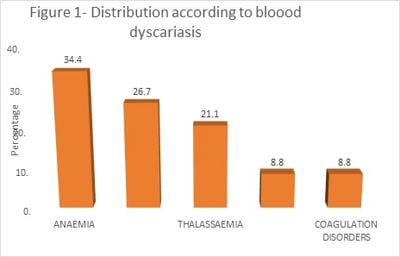
Background: We aimed to study the spectrum of ophthalmic manifestations in blood dyscrasias and assess the prevalence of ocular manifestations between acute and chronic blood malignancies. The study also aimed to correlate ophthalmic findings in anaemia, thalassaemia, leukaemia, lymphomas and other bleeding disorders. Methodology: The study was conducted as an observational study on a total of 180 patients diagnosed with blood dyscrasias at the Department of Ophthalmology, tertiary care hospital, during the study period of 2 years. Detailed history and examination were conducted, and findings were noted. All relevant haematological investigations were also done. Results: Out of 180 cases, anaemia was the most common diagnosis (34.4%). In patients with anaemia, 61.2% of subjects had normal findings, whereas 38.7% of cases had conjunctival pallor. However, in sickle cell anaemia, conjunctival pallor and comma sign was observed in 62.5% of patients each. Retinal Veinous Tortuosity was the most common finding in Thalassemia. In contrast, Veinous Fullness and Tortuosity and sub - conjunctival bleeding were the most common finding in patients with leukemia and coagulation disorders, respectively. Conclusion: Ophthalmic manifestations are commonly observed in patients with blood dyscrasia. The routine ocular examination must be conducted in cases of blood dyscrasias, and especially posterior segment evaluation should be mandatory in severe anaemia/ sickle cell haemo - globinopathy and leukaemia, which is both diagnostic and prognostic of the severity of this disease. Also, there is a need to develop a standard referral protocol between the haematology clinic and the eye clinic so that blood dyscrasia patients can have periodic evaluations.

Keywords: Opthmological manifestations, Blood dyscrasia, Anemia, ITP, Coagulation disorders

Corresponding Author How to Cite this Article To Browse
Shreya Tyagi, Resident, Department of Ophthalmology, Gandhi Medical College, Bhopal, Madhya Pradesh, India.
Email:
Shreya Tyagi, Vivek Som, Kavita Kumar, Rajendra Nigam, Study of ophthalmic manifestations in blood dyscrasias. Trop J Ophthalmol Otolaryngol. 2021;6(5):85-92.
Available From
https://opthalmology.medresearch.in/index.php/jooo/article/view/210

Manuscript Received Review Round 1 Review Round 2 Review Round 3 Accepted
2021-08-21 2021-08-27 2021-09-10 2021-09-19 2021-09-28
Conflict of Interest Funding Ethical Approval Plagiarism X-checker Note
No Nil Yes 8%

© 2021 by Shreya Tyagi, Vivek Som, Kavita Kumar, Rajendra Nigam and Published by Siddharth Health Research and Social Welfare Society. This is an Open Access article licensed under a Creative Commons Attribution 4.0 International License https://creativecommons.org/licenses/by/4.0/ unported [CC BY 4.0].

Introduction

The blood is common to every tissue, and its disorder can affect any part of the body. The ophthalmic symptoms may be initial mode of presentation of a number of blood dyscrasia so the ophthalmologist is often the first witness before the patient reaches the haematologist for final diagnosis. Anemia is the most common haematological disorder in India, which the pallor of palpebral conjunctiva can identify, although not a highly accurate index of severity of anemia. Damage by anemia can be an indicator for retinal damage manifesting as haemorrhage or pallor.[1]. As the disease progresses, haemorrhages, exudates, distended tortuous retinal veins and ultimately even papilloedema may occur in the ocular fundus.[2].

Leukaemia may affect any ocular tissue either by direct infiltration of the leukemic cells or the secondary effect of the neoplasm. Changes in the retina are the most common clinical manifestation of Leukemic involvement of the eye.[2]. These manifestations include vascular sheathing and tortuosity, pallor, haemorrhages and exudates, cotton wool spots and neovascularization at the periphery of the disc.[3]. The presence of ocular involvement is associated with poor prognosis in acute childhood leukaemia.[2]. Coagulative disorders such as Purpuras can present with haemorrhages involving the whole of the retina and vitreous in young girls, especially those who suffer from Idiopathic thrombocytopenic purpura.[4].

Sickle cell disease can present with lid edema, conjunctival sickling sign, iris atrophy, and iris neo-vascularisation and angioid streaks. In the sickle cell, Thalassemia exudative and haemorrhagic changes have been found in the retina[5]. Notably, the incidence of proliferative retinopathy is highest in patients with HbSC or S-beta Thal (33% and 14%, respectively), while patients with HbSS have a 3% incidence of proliferative retinopathy.[6]. Ocular effects in thalassaemia patients include visual acuity changes, cataract, pigmentary retinopathy, optic neuropathy, thinning and tortuosity of retinal vessels, & vitreoretinal haemorrhages.

Ophthalmic manifestations in polycythemia are seen due to an increase in blood viscosity with slowing circulation and resulting hypoxia. The changes may be seen from dilated tortuous retinal veins, haemorrhages, papilledema to central retinal vein occlusion. [2].

Various reports indicate that there exists a link between haematological abnormality and ocular manifestations.[7]. Since ophthalmic manifestations in several blood dyscrasias precede the onset of symptoms and in some cases denote the severity of disease, ophthalmic evaluation becomes mandatory in blood dyscrasias. Fundus changes in haematological disorders are studied in the literature. Still, ocular manifestations in haematological disorders as a whole are poorly studied, so we have tried to survey the above facts in detail.

Ophthalmic evaluation can therefore help in understanding the patterns, and appropriate screening protocols can be designed. The present study was therefore conducted to study the spectrum of ophthalmic manifestations in blood dyscrasias and to assess the prevalence of ocular manifestations between acute and chronic blood malignancies. The study also aimed to correlate ophthalmic findings in anaemia, thalassaemia, leukaemia, lymphomas and other bleeding disorders. This will help prevent the ophthalmic changes early during disease and prevent the morbidity and blindness occurring thereof.

Methodology

Setting: Patients with blood dyscrasias referred for complete ophthalmic examination from the Department of Medicine and Paediatrics to Department of Ophthalmology, Gandhi Medical College, and associated Hamidia Hospital, Bhopal

Duration and type of study: 2 years, i.e. from 1st November 2018 to 30th September 2020 and Prospective Observational Study

Sampling methods: Purposive sampling

Sample size: 180 patients diagnosed with blood dyscrasias.

Inclusion criteria: All the patients diagnosed with blood dyscrasias, including anaemia, thalassaemia, leukaemia and bleeding disorders; belonging to an age group of fewer than 70 years and giving consent for the complete ophthalmic examination

Exclusion criteria: whereas pregnant females; patients with the known ocular disorder; with a history of any ocular trauma due to RTA/head injury/ocular injury and not giving consent for the study.


Data collection procedure: Detailed history regarding sociodemographic variables such as age, gender, socioeconomic status etc., was obtained and entered the questionnaire. Clinical history regarding the blood dyscrasias, presence of ocular symptoms, time since diagnosis, chemotherapy received, and other relevant information was obtained from all the study participants and documented. All the patients were then subjected to detailed general and systemic examination. The examination of the eyes was carried out at the bedside or in the eye department. Visual acuity was recorded using Snellen charts, and refractive error was assessed.

Anterior segment examination was done using a slit-lamp. Intrao - ocular pressure was measured using schiotz tonometer and applanation tonometer. Fundus examination was done by slit-lamp biomicroscopy, binocular indirect ophthalmoscopy. Fundus Fluorescein angiography and fundus photography were done wherever indicated. Gonioscopy (by indirect three mirrored gonio lens) was done wherever indicated. Additional procedures including Fundus Fluorescein Angiography (FFA) and OCT, B- SCAN of the eyes, central nervous system examination were done in cases suggested.

Further, all relevant hematological investigations like Hb%, TLC, DLC, peripheral smear, reticulocyte count, serum iron studies, bleeding and coagulation profile, serum iron studies were done in all the cases. Other routine investigations like serum electrolytes, urine routine microscopy, blood sugar profiles were ordered wherever indicated. And appropriate hematological/ histopathological investigations like bone marrow studies were ordered whenever indicated. Documentation of ophthalmological pathological, haematological and histopathological findings was done in a preformed Proforma.

Ethical consideration & permission: The study was approved by the ethical committee .

Statistical Analysis: The collected data was entered in MS excel sheet, and the data were analysed using IBM SPSS software version 20. Categorical data were expressed as frequency and percentage, whereas numerical data were expressed as mean and standard deviation.

Results

A total of 180 patients were included in our study with various blood ascariasis.

Out of 180 patients, anemia was the most common diagnosis observed in 62 (34.4%) cases, followed by sickle cell anemia (26.7%). (Figure 1) Out of 62 patients with anemia, the majority of cases, i.e. 27 (42.5%), had iron deficiency anemia followed by 12 (19.3%) and 5 (8%) cases with megaloblastic and aplastic anemia, respectively. However, anemia secondary to other causes was observed in 18 (29%) cases. Out of 48 patients with sickle cell anemia, Sickle Cell Anaemia (Homozygous) Hb-SS was seen in 24 (50%), Sickle Cell Trait (SC-As) in 16 (33.3%), Sickle Cell Disease-Thalassemia (Sc-Thal) in 6 (12.5%) and HbD (Punjab variant) were noted in 2 (4.1%) cases. Among 16 patients, AML, ALL, CML and CLL were observed in 4, 3, 8 and 1 cases, respectively.

opthal_210_01.jpg

Table 1- Distribution according to sociodemographic variables

Sociodemographic variables No. of patients (n=180) Percentage
Gender Male 93 51.7
Female 87 48.3
Age (years) 0-10 52 28.9
10-20 39 21.7
20-30 40 22.2
30-40 22 12.2
40-50 16 8.9
50-60 8 4.4
>60 3 1.7

The majority of cases were males in our study (51.7%). The mean age of patients was 27.4 years, and the majority of patients belonged 0 to 10 years (28.9%), followed by 30 to 40 years (22.2%). (Table 1)

Table 2- Ophthalmic manifestations in various blood dyscrasias

Blood dyscrasias Ophthalmic finding No. of patients Percentage
Anemia (n=62) Normal 38 61.2%
Conjunctival Pallor 24 38.7%

  The pallor of fundus, Veinous Tortuosity/venous Fullness 23 37%
Flame Shaped Haemorrhages With White Centres 18 29%
Superficial Haemorrhages With Cotton Wool Spots 6 9.6%
Neo- Vascularisation 3 4.8%
Subhyaloid Haemorrhage 2 3.2%
Sickle cell anemia (n=48) Conjunctival Pallor 30 62.5%
Conjunctival Sign (Comma Sign) 30 62.5%
Normal 18 37.5%
The pallor of Fundus with Tortuosity of Retinal Veins 30 62.5%
Flame Shaped Haemorrhages 24 50%
Cotton Wool Spots 13 27.08%
Retinitis Proliferans with Neovascularisation 2 4.1%
Black Sunburst Sign 1 2.0%
Vitreous Haemorrhage 2 4.1%
Thalessemia (n=38) Normal 18 47.3%
Dry Eye 3 5.2%
Lenticular Opacities 2 13.1%
Retinal Veinous Tortuosity 12 31.5%
Retinal Pigment Epithelium Changes(Degeneration/Mottling) 10 26.31%
Defective Color Vision 2 5.2%
Defective Visual Field 1 2.6%
Leukemia (n=16) Normal 4 25%
Orbital Involvement 1 6.2%
Sub-Conjunctival Haemorrhage 1 6.2%
Veinous Fullness and Tortousity 12 75%
Flame Shaped Haemorrhage with White Centres 8 50%
Cotton Wool Spots 2 12.5%
Pre-Retinal Haemorrhage 4 25%
Papilloedema with Neo-Vascularisation Over Disc 2 12.5%
Coagulation disorders (n=16) Normal 9 56.25%
  Lid Edema 2 12.5%
Sub- Conjunctival Haemorrhage 6 37.5%
Veinous Fullness/ Tortuosity 5 31.25%
Retinal Haemorrhage (Flame- Shaped/Sub Hyaloid/Peri-Papillary) 7 43.75%
Cotton Wool Spots 3 18.75%
Retinal Detachment 2 12.5%
Disc Changes (Papilloedema) 2 12.5%

Table 2 reveal various findings in patients with blood dyscrasias. In patients with anemia, 61.2% of cases had normal results, whereas 38.7% had conjunctival pallor. However, in sickle cell anemia, conjunctival pallor and comma sign was observed in 62.5% of cases each. Retinal Veinous Tortuosity was the most common finding in Thalassemia. In contrast, Veinous Fullness and Tortuosity, and subconjunctival hemorrhage were the most common findings in patients with leukemia and coagulation disorders, respectively.

Table 3- Ocular manifestations in acute and chronic leukemia

Ocular findings (n=16) AML ALL CML CLL TOTAL
Normal 1   3   4
Orbital Involvement 1       1
Sub-Conjunctival Haemorrhage 1       1
Veinous Fullness and Tortousity 3 3 5 1 12
Flame Shaped haemorrhage with White Centres 2 2 3 1 8
Cotton Wool Spots 1   1   2
Pre-Retinal Haemorrhage 1 2 1   4
Papilloedema with Neo-Vascularisation over Disc   1 1   2
Total cases 4 3 8 1 16

Veinous Fullness and Tortuosity was the most common finding in all types of leukemia, whether acute or chronic leukemia. (Table -3).


Table 4- Frequency of ophthalmic findings in blood dyscrasias: a comparative review

Ophthalmic Findings- Posterior Segment Total N= 180 Anaemia Sickle Cell Anaemia Thalassaemia Leukaemia Coagulative Disorders
Pallor of Fundus 84(46.6%) 23(12.7%) 30(16.6%) 12(6.6%) 12(6.6%) -
Veinous Tortuosity/ Fullness 84(46.6%) 23(12.7%) 30(16.6%) 12(6.6%) 12(6.6%) 7(3.8%)
Retinal Haemorrhages 54(30%) 18(10%) 24(13.3%) - 8(10%) 4(2.2%)
Cotton Wool Spots 24(13.3%) 6(3.3%) 13(7.2%) - 2(1.1%) 3(1.6%)
Neo- Vascularisation 9(5%) 3(1.6%) 2(1.1%) - 4(2.2%) -
PreRetinal Haemorrhage 6(3.3%) 2(1.1%) 2(1.1%) - 2(1.1%) 4(2.2%)
Vitreous Haemorrhage 6(3.3%) 2(1.1%) 2(1.1%) - 2(1.1%) 4(2.2%)

The above table suggests that the most common findings in blood dyscrasias were pallor of the fundus(46.6%) and veinous tortuosity/ fullness (46.6%). Both these findings were observed in maximum proportions of cases with anemia, sickle cell anemia and Thalassemia. However, retinal hemorrhages were the most common finding in leukemia, whereas Veinous Tortuosity/ Fullness was the most common finding in coagulation disorders.

Discussions

The ophthalmic symptoms in patients with blood dyscariasis may be the initial presentation and may represent the severity of illness. There were 180 different cases of different blood dyscrasias, and among them, 62 patients (34.4%) were of anaemia. All the possible causes of anaemia were included. About 16 cases (8.8%) were diagnosed as leukaemia, 38 patients with Sickle cell haemoglobinopathy were studied.

Anemia: The maximum number of patients had iron deficiency anemia (43.54%) which is a common factor in our country. Anemia was observed in patients with a mean age of 25.11 years with a slightly high male: female ratio (1.38:1). Iron deficiency anemia was most commonly observed among females, while aplastic anemia was most common in males.

Table 5- Comparison of most common manifestations of blood dyscrasia

Study Nutritional anemia Age Groups Affected (Years) Most Common Ocular Features
Merin et al[8] 30% 20- 40 Retinal Haemorrhages (55%)
Holt et al[9] 49% 40-60 Retinal Haemorrhages (67%)
Rubenstein et al[10] 22% 20-50 Retinal Haemorrhages (41%)
Lange et al[1] 37% 25-60 Pallor of Fundus (70%)> Retinal Haemorrhages
Our Study 43.54% 20-40 Pallor of Fundus (37%)> Retinal Haemorrhages (29%)

In our study, 39% of cases with anemia had retinopathy. Similarly, 28.35% and 22.5% of patients had retinopathy in a survey by Rubenstein et al. [10] and Merin et al. [8], respectively. Some signs were common to every case, viz., the pale background of the fundus, venous fullness; axial reflex in both arteries and veins were reduced in brilliance.

The subsequent common finding comprised of flame-shaped haemorrhages and haemorrhages with central pallor suggestive of Roth spots. (29%). 9.6% of the cases showed superficial haemorrhages with cotton wool exudates. In 2 cases, sub- hyaloid hemorrhage was found. Vitreous hemorrhage was found in 2 cases, one with haemoglobin 5gm/dl and 3 gm/dl. All cases of anaemia with fundus findings were with haemoglobin 7.5 % or less. It has been documented in the literature that fundus findings have been associated with a profound fall of haemoglobin, and a critical level of 50% fall has been described by Ballantyne et al.[6].

Sickle Cell Anaemia: Out of 48 patients with sickle cell anemia, 30(62.5%) patients had conjunctival sign positive in them, and 20 of them belonged to SS, had AS, and 4 were cases of sickle- thalassemia trait. Seargent et al. [11]. reported positive conjunctival signs in 32 out of 38 patients, 4 in patients of SC and 1 out of AS patients. Cordon et al. [12] reported corkscrew-like dilated conjunctival vessels in 74 out of 76 SS patients. They correlated this finding with irreversibly sickled cells. Though varying incidence in various studies has been observed, it is agreed that dilatation and tortuosity of the blood vessels are one of the commonest and earliest findings in sickle cell retinopathy. Black Sunburst Sign was seen in 1 patient (2%) of SS with the recurrent occlusive crisis. Condon et al [12]. reported this sign in 32.9% of sickle cell disease cases.

Our study observed retinitis proliferates with neovascularization in 2 cases, and two patients with vitreous hemorrhages were seen—similarly, Condon et al. [12]. The authors described retinitis proliferans to be common in HbC (SC) and auto- infarction in homozygous (SS) disease. Meurs et al. [13] observed proliferative retinopathy in 2% of SS patients and 50% of SC patients. It led to vitreous hemorrhage in 18% and RD in 8% of cases.

Leukaemia: In this study, 16 cases with leukaemia were enrolled, of which chronic leukaemia was found more in number [9] than acute leukaemia[7]. About 75% of subjects had venous fullness and tortuosity, 50% of patients had flame-shaped hemorrhage with white centres, 25% of patients had pre retinal hemorrhage. However, 12.5% had neovascularisation on the disc and cotton wool spots in the general fundus, and 12.5% had bilateral papilloedema. Orbital involvement and Subconjunctival hemorrhage were seen in 6.2% of cases each.


Retinopathy in leukemia was observed in 56% of Kataria et al. [14] and 44% cases by Holt et al. [8]. The higher incidence of retinopathy in leukemia in our study compared to other studies could be because leukaemia patients with severe disease were admitted and referred for their ophthalmic examination.

Retinal hemorrhages are extremely common in the leukemic fundus, more common in the acute type of the disease. They may be only and the most frequent and severe ocular complication[15]. They are usually flame-shaped, small and scattered, occurring preferentially near the posterior pole and typically have a central white area. Superficial flame-shaped hemorrhages with white centres were found in 78.57% of cases in the present study.

They were large sometimes. Sometimes deep hemorrhages were also present, mainly at the posterior pole. In one patient with acute myeloid leukemia along with superficial hemorrhages, Cotton wool spots were found. Retinal hemorrhages do not always indicate severe anaemia, and that severe anaemia inpatient with leukemia does not necessarily entail retinal hemorrhage. Ballantype et al. [6] suggested that the centre represented the accumulations of leukocytes.

Thalassaemia: Most common ocular finding in patients with Thalassemia was retinal veinous tortuosity (31.5%) followed by retinal pigment epithelium changes (26.3%). Colour vision was impaired in 5.2%, whereas lenticular opacity and visual field defects could be observed in 13.1% and 2.6% cases, respectively. Similar findings were observed by Jafari et al. [16] in which ocular findings such as the dry eye (33.3%), cataract (10.2%), retinal pigment epithelium degeneration (16.7%), colour vision deficiency (3.7%) and visual field defects (33.7%) were detected in 68.5% of the thalassemic group. Barteselli et al [17]. reported ocular fundus abnormalities characteristic of pseudoxanthoma elasticum (PXE) in 70 of 255 patients (27.8%) with Thalassemia. Taneja et al [18]. However, documented Retinal Pigment Epithelium (RPE) degeneration in 15/45 patients and RPE mottling in 4/45 patients with Thalassemia.

Coagulation Disorders: Most of the patients with coagulation disorders in our study had normal fundus findings (56%). In the anterior segment, the most common finding observed was sub - conjunctival hemorrhage.

These findings were supported by Sodhi et al. [19], in which the authors stress subconjunctival bleeding as the first presenting clinical feature in ITP. Retinal hemorrhages were noted in 43.75 % of the cases in our study. This was in agreement with several studies conducted in which intraocular hemorrhages including subretinal, intraretinal, subhyaloid and vitreous hemorrhages were common.[7,20].

Conclusion

Ophthalmic manifestations are commonly observed in patients with blood dyscrasia. The routine ocular examination must be conducted in blood dyscrasias; especially posterior segment evaluation should be mandatory in severe anaemia/ sickle cell haemoglobinopathy and leukaemia, which is both diagnostic and prognostic of the severity of this disease. Also, there is a need to develop a standard referral protocol between the haematology clinic and the eye clinic so that blood dyscrasia patients can have periodic evaluations. The signs noted during ophthalmic manifestations of blood dyscrasias reveal that they can be severe (vitreous hemorrhage, retinal detachment, sub- hyaloid hemorrhage etc.) and involve the functional prognosis of the eye, especially in young patients. Their insidious evolution is always a cause of irreversible blindness, especially in our regions where delays in presentation and diagnosis are deplored. The work has been done by four authors, each having a crucial part to play.

Author contribution

Dr. Shreya collected data and wrote the manuscript.

Dr. Vivek Som sir helped compile it and organise it into discernible tables and gave guidance regarding the writing of the manuscript.

Dr. Kavita Kumar ma’am helped in inter-departmental support and easy reference and communication between the different specialities (department of medicine, paediatrics and pathology). She has been a guiding source throughout.

Dr. R. Nigam, sir, helped understand the blood dyscrasias and haematological parameters better and also provided information of patients’ different investigations and helped segregate patients of different blood dyscrasias.


What does this study add to existing knowledge?

  1. This study screens a large group of patients with blood dyscrasias in an institute that has an inflow of patients of every blood disorder and of every age group.
  2. Ophthalmic manifestations in blood dyscrasias are more common than realised.
  3. Leukaemia (acute and chronic) were found to have involvement in about 75% of the patients, which is higher than other studies.
  4. Posterior segment involvement was maximum in leukaemia(75%), followed by sickle cell anaemia(62.5%).
  5. Sight threatening complications like vitreous haemorrhage, retinal detachment, sub- hyaloid haemorrhage etc. are seen commonly (5.5% of the patients in this study) in patients with blood dyscrasias.

Reference

01. Lang GE, Spraul CW, Lang GK. Okuläre Veränderungen bei hämatologischen Grunderkrankungen [Ocular changes in primary hematologic diseases]. Klin Monbl Augenheilkd. 1998 June;212(6):419-27. German. doi: 10.1055/s-2008-1034925 [Crossref][PubMed][Google Scholar]

02. Pandharinath, Jakkal Tapan, Shitole Satish Chandrakant, and Jakkal Darpan Pandharinath. Ophthalmic Manifestations Of Common Haematological Disorders. Journal of Evolution of Medical and Dental Sciences. 3;42(2014): 10510-10517. [Crossref][PubMed][Google Scholar]

03. Delaney WV Jr, Kinsella G. Optic disk neovascularization in leukemia. Am J Ophthalmol. 1985 Feb 15;99(2):212-3. doi: 10.1016/0002-9394(85)90238-7 [Crossref][PubMed][Google Scholar]

04. Srinivasan M, Chakrabarti A, Chakrabarti M. Expulsive haemorrhage in a case of thrombocytopenic purpura. Indian J Ophthalmol. 1996 Mar;44(1):44-5. [Crossref][PubMed][Google Scholar]

05. Duke - Elder, John H. Dobree: Diseases of the retina. System of Ophthalmology. 1967, Vol 10: pp 376-99. [Crossref][PubMed][Google Scholar]

06. Ballantyne, Arthur James, and Isaac Chesar Michaelson. Textbook of the Fundus of the Eye. Churchill Livingstone. 1970. [Crossref][PubMed][Google Scholar]

07. Majji AB, Bhatia K, Mathai A. Spontaneous bilateral peripapillary, subhyaloid and vitreous hemorrhage with severe anemia secondary to idiopathic thrombocytopenic purpura. Indian J Ophthalmol. 2010 May-Jun;58(3):234-6. doi: 10.4103/0301-4738.62651 [Crossref][PubMed][Google Scholar]

08. Merin S, Freund M. Retinopathy in severe anemia. Am J Ophthalmol. 1968 Dec;66(6):1102-6. doi: 10.1016/0002-9394(68)90818-0 [Crossref][PubMed][Google Scholar]

09. Holt JM, Gordon-Smith EC. Retinal abnormalities in diseases of the blood. Br J Ophthalmol. 1969 Mar;53(3):145-60. doi: 10.1136/bjo.53.3.145 [Crossref][PubMed][Google Scholar]

10. Rubenstein RA, Yanoff M, Albert DM. Thrombocytopenia, anemia, and retinal hemorrhage. Am J Ophthalmol. 1968 Mar;65(3):435-9. doi: 10.1016/0002-9394(68)93098-5 [Crossref][PubMed][Google Scholar]

11. Serjeant GR, Serjeant BE, Condon PI. The conjunctival sign in sickle cell anemia, A relationship with irreversibly sickled cells. JAMA. 1972 Mar 13;219(11):1428-31. [Crossref][PubMed][Google Scholar]

12. Condon PI, Serjeant GR, Ikeda H. Unusual chorioretinal degeneration in sickle cell disease, Possible sequelae of posterior ciliary vessel occlusion. Br J Ophthalmol. 1973 Feb;57(2):81-8. doi: 10.1136/bjo.57.2.81 [Crossref][PubMed][Google Scholar]

13. van Meurs JC. Visusbedreigende oogafwijkingen bij patiënten met sikkelcelziekte op Curaçao [Vision-threatening eye manifestations in patients with sickle cell disease on Curaçao]. Ned Tijdschr Geneeskd. 1990 Sep 15;134(37):1800-2. Dutch [Crossref][PubMed][Google Scholar]

14. Kataria VC, Audich KL, Narang SK, Khamar BM. The fundus findings in blood dyscrasias. Indian J Ophthalmol. 1983;31 Suppl:899-902. [Crossref][PubMed][Google Scholar]


15. Reddy SC, Jackson N, Menon BS. Ocular involvement in leukemia--a study of 288 cases. Ophthalmologica. 2003 Nov-Dec;217(6):441-5. doi: 10.1159/000073077 [Crossref][PubMed][Google Scholar]

16. Jafari R, Heydarian S, Karami H, Shektaei MM, Dailami KN, Amiri AA, et al. Ocular abnormalities in multi-transfused beta-thalassemia patients. Indian J Ophthalmol. 2015 Sep;63(9):710-5. doi: 10.4103/0301-4738.170986 [Crossref][PubMed][Google Scholar]

17. Barteselli G, Dell'arti L, Finger RP, Charbel Issa P, Marcon A, Vezzola D, et al. The spectrum of ocular alterations in patients with β-thalassemia syndromes suggests a pathology similar to pseudoxanthoma elasticum. Ophthalmology. 2014 Mar;121(3):709-18. doi: 10.1016/j.ophtha.2013.10.016 [Crossref][PubMed][Google Scholar]

18. Taneja R, Malik P, Sharma M, Agarwal MC. Multiple transfused thalassemia major: ocular manifestations in a hospital-based population. Indian J Ophthalmol. 2010 Mar-Apr;58(2):125-30. doi: 10.4103/0301-4738.60083. PMID: 20195035; [Crossref][PubMed][Google Scholar]

19. Sodhi PK, Jose R. Subconjunctival hemorrhage: the first presenting clinical feature of idiopathic thrombocytopenic purpura. Jpn J Ophthalmol. 2003 May-Jun;47(3):316-8. doi: 10.1016/s0021-5155(03)00017-0 [Crossref][PubMed][Google Scholar]

20. Turaka K, Shields CL, Bianciotto C, Shields JA. Vitreous hemorrhage as the initial manifestation of idiopathic thrombocytopenic purpura. Retin Cases Brief Rep. 2012 Winter;6(1):16-8. doi: 10.1097/ICB.0b013e3181f7f75d [Crossref][PubMed][Google Scholar]