E-ISSN:2456-6454
P-ISSN:2581-4907
RNI:MPENG/2017/74152

Case Report

Retinochoroidal coloboma

Tropical Journal of Ophthalmology and Otolaryngology

2021 Volume 6 Number 5 September October
Publisherwww.medresearch.in

Unusual Co-existence of Optic disc pit and Retino-choroidal coloboma- Case report

Priya A.1*, Anusha T.2
DOI: https://doi.org/10.17511/jooo.2021.i05.04

1* Aneesha Priya, Resident, Department of Ophthalmology, Santhiram Medical College and General Hospital Nandyal, Kurnool, AP, India.

2 T. Anusha, , Department of Ophthalmology, Katuri Medical College, Guntur, AP, India.

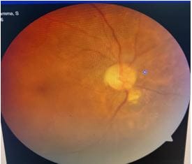
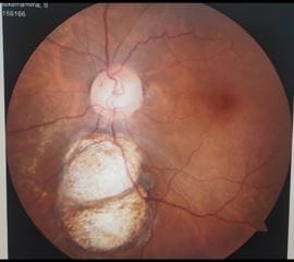
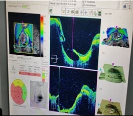
The co-existence of optic disc pit with Retinochoroidal coloboma is rare. Only a few cases of optic disc pit co-occurring with serous macular detachment, choroidal coloboma or retinochoroidal coloboma were reported. We report a case of Optic pit with Retinochoroidal coloboma without macular involvement. A 50-year-old woman presented to our clinic for a routine bilateral ophthalmologic examination. Her BCVA was 6/12 in the right eye and 6/9 in the left eye. Fundus examination of the right eye revealed aborted coloboma inferior to the optic disc. In the left eye, there was an oval-shaped and grey temporal optic disc pit and a Retino-choroidal coloboma low to the optic disc not involving disc or macula. Macula was normal in both eyes. The patient was monitored regularly for possible complications like retinal detachment or neovascularization.

Keywords: Retino choroidal Coloboma, Aborted coloboma, Optic disc pit

Corresponding Author How to Cite this Article To Browse
Aneesha Priya, Resident, Department of Ophthalmology, Santhiram Medical College and General Hospital Nandyal, Kurnool, AP, India.
Email:
Aneesha Priya, T. Anusha, Unusual Co-existence of Optic disc pit and Retino-choroidal coloboma- Case report. Trop J Ophthalmol Otolaryngol. 2021;6(5):105-107.
Available From
https://opthalmology.medresearch.in/index.php/jooo/article/view/209

Manuscript Received Review Round 1 Review Round 2 Review Round 3 Accepted
2021-09-07 2021-09-09 2021-09-16 2021-09-23 2021-09-30
Conflict of Interest Funding Ethical Approval Plagiarism X-checker Note
Nil Nil Yes 16%

© 2021by Aneesha Priya, T. Anushaand Published by Siddharth Health Research and Social Welfare Society. This is an Open Access article licensed under a Creative Commons Attribution 4.0 International License https://creativecommons.org/licenses/by/4.0/ unported [CC BY 4.0].

Introduction

Optic disc pit is usually a congenital anomaly of optic nerve head but acquired cases seen as secondary to glaucoma or myopia [1]. They are present along the rim of the optic disc and commonly cause serous detachments of the macula, associated with retinal pigment epithelium mottling and cystic changes [2]. It occurs unilaterally and temporally on the optic disc but can be seen centrally or anywhere on the margin of the optic disc [3]. They are hypopigmented, round or oval, greyish and may co-exist with the cilioretinal artery. They are asymptomatic and can be detected incidentally. Ocular coloboma occurs as an isolated defect in healthy individuals or part of a complex malformation syndrome. Optic disc pits are typically not associated with iris or retino-choroidal coloboma. Here we described a rare case of optic disc pit co-occurring with retino-choroidal coloboma.

Case report

A 50-year-old woman presented to our clinic for a routine bilateral ophthalmologic examination. Her BCVA was 6/12 in the right eye and 6/9 in the left eye. Intraocular pressure was 13 mmHg in the right eye and 12 mmHg in the left eye. The right eye lens was cataractous, and the left eye was pseudophakic. Fundus examination of the right eye with 0.4 CDR revealed aborted coloboma inferior to the optic disc and left eye with 0.6 CDR. There was an oval-shaped and grey temporal optic disc pit and a Retino-choroidal coloboma inferior to the optic disc not involving disc or macula. Macula was normal in both eyes. The patient was monitored regularly for possible complications like retinal detachment or neovascularization.

opthal_209_01.JPG

Figure 1: Fundus photo of the right eye showing Aborted coloboma inferior to Optic disc

opthal_209_02.JPG

Figure 2: Left eye fundus photo with Temporal Optic pit and Retino-choroidal coloboma inferior to Optic disc

opthal_209_03.JPG

Figure 3: (LE) OCT picture showing RetinoChoroidal coloboma

Discussion

Congenital optic disc pits are usually unilateral but can be bilateral in 15% of the population. The embryological basis of congenital pit formations was hypothesized to be due to developmental anomaly of the primordial optic nerve or incomplete closure of the embryonic fissure [4,5,6]. The co-existence of optic disc pits and uveal tissue colobomas supports the latter hypothesis. Incomplete closure of optic fissure affects the developing optic cup or stalk and the adult derivations of these structures, resulting in an inferonasal defect in the optic disc, retina, ciliary body or iris. Colobomas affecting the sensory retina and RPE also involve the choroid because its differentiation depends on the intact RPE layer. Bare sclera is seen with retinal vessels passing over the coloboma cases of optic disc pit co-occurring with serous macular detachment, choroidal coloboma or retinochoroidal coloboma were reported, both unilaterally and bilaterally[5,6]. Visual loss occurs due to leakage


from the optic disc pit and exudation of fluid beneath the macula. In this case report, we described the co-existence of the optic pit with retino choroidal coloboma without macular involvement. Only a few cases were published regarding optic pits and choroidal colobomas [5,6].

Conclusion

Taking the basis of these case reports, a defect in the closure of the embryonic fissure appears to be the possible etiology of the optic pit—regular follow-up of the patient with spectral-domain. OCT is required for monitoring macular involvement.

Reference

01. Healey PR, Mitchell P. The prevalence of optic disc pits and their relationship to glaucoma. J Glaucoma. 2008 Jan-Feb;17(1):11-4. doi: 10.1097/IJG.0b013e318133fc34 [Crossref][PubMed][Google Scholar]

02. Kalogeropoulos D, Ch'ng SW, Lee R, Elaraoud I, Purohit M, Felicida V, et al. Optic Disc Pit Maculopathy: A Review. Asia Pac J Ophthalmol (Phila). 2019 May-Jun;8(3):247-255. doi: 10.22608/APO.2018473 [Crossref][PubMed][Google Scholar]

03. Ohno-Matsui K, Hirakata A, Inoue M, Akiba M, Ishibashi T. Evaluation of congenital optic disc pits and optic disc colobomas by swept-source optical coherence tomography. Invest Ophthalmol Vis Sci. 2013 Nov 25;54(12):7769-78. doi: 10.1167/iovs.13-12901 [Crossref][PubMed][Google Scholar]

04. Gass JD. Serous detachment of the macula. Secondary to congenital pit of the optic nervehead. Am J Ophthalmol. 1969 Jun;67(6):821-41. doi: 10.1016/0002-9394(69)90075-0 [Crossref][PubMed][Google Scholar]

05. Brown GC, Augsburger JJ. Congenital pits of the optic nerve head and retinochoroidal colobomas. Can J Ophthalmol. 1980 Jul;15(3):144-6. [Crossref][PubMed][Google Scholar]

06. Saatci, Ali Osman, Nilufer Kocak, and Fatma Meltem Soylev. "Unilateral coexistent optic pit and choroidal coloboma. " Neuro-ophthalmology 28. 1 (2004): 41-43. [Crossref][PubMed][Google Scholar]