E-ISSN:2456-6454
P-ISSN:2581-4907
RNI:MPENG/2017/74152

Research Article

Hematoma Auris

Tropical Journal of Ophthalmology and Otolaryngology

2021 Volume 6 Number 5 September October
Publisherwww.medresearch.in

Window technique for Hematoma Auris / Seroma – A Promising technique

Wahid S.1, Susmitha S.2*, Yedluri S.3, Dinesh A.4
DOI: https://doi.org/10.17511/jooo.2021.i05.03

1 Sk Wahid, Assistant Professor, Department of Otorhinolaryngology, NRI Academy of Sciences, Guntur, Andhra Pradesh, India.

2* Satujoda Susmitha, Post Graduate, Department of Otorhinolaryngology, NRI Academy of Sciences, Guntur, Andhra Pradesh, India.

3 Satya Prabhakar Rao Yedluri, Professor and HOD, Department of Otorhinolaryngology, NRI Academy of Sciences, Guntur, Andhra Pradesh, India.

4 Aradhyula Dinesh, Post Graduate, Department of Otorhinolaryngology, NRI Academy of Sciences, Guntur, Andhra Pradesh, India.

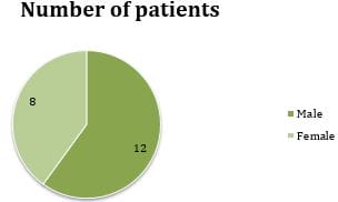
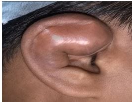
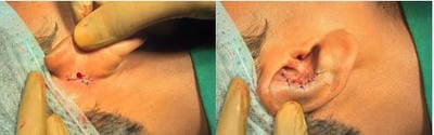
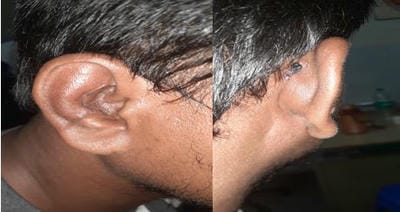
Background: The seroma of the pinna is the accumulation of serous fluid between the perichondrium and cartilage of the pinna. Despite several surgical techniques in the treatment of seroma, recurrence is common. The aim and objective of this preliminary study are to present and discuss a novel approach in treating pinna's pseudocyst. Materials and methods: A total of 20 patients with auricular seromas treated by window technique were included in the current study. Results: Results showed that all the patients tolerated the procedure well. No patient had any collection of fluid after the removal of the dressing. No patient experienced pain, fever, or oedema after the treatment. The seroma disappeared without disfigurement. There were no recurrences on further follow‑up. Conclusion: The window technique is a valuable technique for treating seroma and hematoma of pinna as the outcome is good with no recurrence.

Keywords: Hematoma Auris, Perichondrium, Seroma, Pinna, Cartilage, Serous fluid, Window technique, Pseudocyst

Corresponding Author How to Cite this Article To Browse
Satujoda Susmitha, Post Graduate, Department of Otorhinolaryngology, NRI Academy of Sciences, Guntur, Andhra Pradesh, India.
Email:
Sk Wahid, Satujoda Susmitha, Satya Prabhakar Rao Yedluri, Aradhyula Dinesh, Window technique for Hematoma Auris / Seroma – A Promising technique. Trop J Ophthalmol Otolaryngol. 2021;6(5):99-104.
Available From
https://opthalmology.medresearch.in/index.php/jooo/article/view/201

Manuscript Received Review Round 1 Review Round 2 Review Round 3 Accepted
2021-10-07 2021-10-09 2021-10-16 2021-10-23 2021-10-30
Conflict of Interest Funding Ethical Approval Plagiarism X-checker Note
Nil Nil Yes 17%

© 2021by Sk Wahid, Satujoda Susmitha, Satya Prabhakar Rao Yedluri, Aradhyula Dineshand Published by Siddharth Health Research and Social Welfare Society. This is an Open Access article licensed under a Creative Commons Attribution 4.0 International License https://creativecommons.org/licenses/by/4.0/ unported [CC BY 4.0].

Introduction

Pseudocyst of the pinna or auricular seroma is a benign cystic swelling with the collection of serous fluid between the perichondrium and cartilage [1]. and does not have an epithelial lining. [1,2]. The scaphoid fossa is involved in most patients, followed by the triangular fossa and cymba concha [3]. Aspiration from the pseudocyst produces a viscous, oily, sterile, pale yellow coloured fluid with a negative bacterial culture[4].

Demography: Males are commonly affected, and the mean age of presentation is 35-40 years.

Etiology: The exact cause for pseudocyst development is unknown though some say it is due to a defect in the embryogenesis. In contrast, others attribute it to repeated trauma or following surgery of pinna. Repeated minor trauma like sleeping on hard pillows, carrying large weights on the shoulder may contribute to the development of a pseudocyst.

Extravasated blood might clot, leading to the deformity of the cartilage as well as the ear. According to the studies of Hansen and Engel, males are usually affected with a unilateral presentation [5]. Other morbidities include scarring, perichondritis, and abscess formation.

Pathology: Some pseudocysts fluids have elevated levels of serum lactic dehydrogenase (L.D.H.) also. LDH-4 and LDH-5 may be released from auricular cartilage degenerated from repeated minor trauma. One article reports that pseudocysts can be regarded as simply a variation of otohematoma or otoseroma. [6]

Diagnosis: Diagnosis is based on clinical characteristics and no evidence of infection. The differential diagnosis of this condition includes cellulitis, relapsing polychondritis, chondrodermatitis helicis, and subperichondrial hematoma secondary to trauma.[7].

Treatment: Procedures like repeated aspiration, incision and drainage with pressure dressing, insertion of drainage tubes, excision, and B.I.P.P. packing can be used as treatment options [8,9,10]. Excision and drainage is the most accepted treatment option, which is the window method where drainage of the collection is done along with removing a piece of cartilage and

perichondrium [11]. Excision of the cartilage and perichondrium can cause complications like scarring, deformity of the pinna, chances of perichondritis and sometimes even an abscess formation. Successful treatment is challenging because of high rates of recurrence. Few researchers have stressed the cosmetic impact of the dressing on the patient, as most patients tend to remove or manipulate the dressing to avoid social embarrassment. Eventually, most of them would return with a recollection of the seroma, leading to a window operation as a second management line. More invasion treatment options have a higher risk of complications like cauliflower-like deformity of the auricle [12].

Aim of the study

This study aimed to access the role of window technique in treating primary or recurrent seroma and knowing the outcomes like recurrence rate, cosmetic outcome and chances of perichondritis/ abscess formation.

Material and Methods

Setting: This study was conducted in N.R.I. medical college(N.R.I.M.C.), a tertiary hospital with all super speciality services available.

Duration and type of the study: This study was conducted on patients with the seroma of the pinna in the department of otorhinolaryngology or N.R.I.M.C. for eight months between August 2019 and March 2020. This is a prospective, interventional study in which window technique surgery was done on patients who presented with the seroma of pinna during the study period.

Sample size calculation: About 20 cases attending the O.P.D. during the eight months were taken into the study after obtaining their informed consent.

Approximately 50% (prevalence) of patients have undergone window technique

Surgery who presented with seroma of pinna at N.R.I.M.C. during the study period. Total 21 patients attended the department with seroma of the pinna. So, the sample size is calculated as per the below formula:

N=z 2 pq/e 2

N=Population size-21


Z: confidence level of 95%-standard value of 1.96

P-expected prevalence or proportion: p=0.5 (since prevalence is 50%)

e- Allowable error-1%

Based on the above formula, the approximate sample size was found to

be 20.

The sample size was calculated after checking the book "A practical

approach to P.G. dissertation" by R Raveendra, Geentanjali et al.

(J.I.P.M.E.R.)

Inclusion Criteria:

Following patients were included in this study:

  • Patients who presented with Primary seroma
  • Patients with recurrent seroma who got seromas previously treated with other options like incision and drainage.
  • Both genders
  • 11-60 years of age

Exclusion criteria:

  • Pregnant & lactating women
  • Patients with severe liver, renal, heart disorders

Methodology or procedure: All the investigations before giving local anesthesia were done on all patients. The procedure was done under strict aseptic conditions. Pinna was anesthetized using 2% xylocaine and adrenaline in 1: 75000 dilution. Infiltration was given in the preauricular and postauricular areas to anesthetize Greater auricular nerve and auriculotemporal nerve. 5 ml of local anaesthetic was used to block the greater auricular and auriculotemporal nerves. A curvilinear incision is given on the depending part of the swelling on the medial aspect of the pinna, and the skin flap is elevated. The corresponding part of the cartilage is removed along with the skin flap on the medial surface. The collection was drained, and the undersurface of the flap was scraped using Rosen's knife followed by pressure dressing for three days. A pressure dressing was applied, and patients were reviewed after one week and then monthly for three months. The outcome was studied regarding recollection, recurrence, development of perichondritis/ abscess and cosmetic outcome.

Materials used: Drug-local anaesthetic-Xylocaine: Xylocaine is an amide local anaesthetic, which is most commonly used for simple surgical and diagnostic procedures which lasts from 30min to 180min[13]. About 2% xylocaine was used along with the adrenaline-the rationale of the combination is that adrenaline acts as a vasoconstrictor which reduces the absorption of xylocaine into the systemic circulation and decreases the blood loss by timely constriction of capillaries. It is also safe to use in end organs where the blood supply is poor[14].

Ethical considerations: Informed consent was taken in the local language from all the study participants. Institutional ethics committee approval was taken before enrolling patients into the study.

Statistical analysis

Frequency or percentage of adverse effects and outcomes were assessed using statistical software named Statistical Package for the Social Science version 20.0.0 (S.P.S.S. Inc., Chicago, Illinois, U.S.A.).

Results

A total of 20 patients with seroma were taken into the study.

Table 1: Type of cases 

Type of cases Number Percentage
Primary 14 70%
Recurrent 6 30%

Out of 20 patients, 14 were primary cases, and six were recurrent cases.

Age:

Table 2: Age distribution of patients

Age group Number Percentage
11-20 years 1 5%
21-30 2 10%
31-40 5 25%
41-50 10 50%
51-60 2 10%

Most of the patients belonged to the age group 41 and 50.

Our study results showed that males have more incidence of seroma compared to females as per various previous studies.


Gender:

Chart 1: Gender distribution of patients

opthal_201_01.JPG

Complications:

Table 3: Complications occurred among study patients

Complication Number of patients Percentage of patients
Recurrence 0 0
Perichondrial reaction 2 10%
Cosmetic deformity 0 0
Pinna thickening 3 15%

Two patients developed perichondrial reaction for which they required treatment with antibiotics and analgesics, and three showed up with pinna thickening with nil recurrence and negligible cosmetic deformity in the three months follow up

opthal_201_02.JPG

Figure 1 : Preop photograph of right ear seroma.

opthal_201_03.JPG

Figure 2 : Intra operative photograph

opthal_201_04.JPG

Figure 3: Post operative follow up period photograph

Comparison with other studies

In the study of Kiran Naik et al. [19], the results showed none of them had any fluid reaccumulation or swelling, similar to our research. One patient had edema of the skin, so the sutures were removed on the 3rd day. Two patients had slight thickening of the aural skin on the 5th day, which returned to normal on the 10th postoperative day. In our study, three patients had a thickening of aural skin, and none reported edema.

The study by harish et al. [20] is a prospective study in which a trans window suture technique was used to treat pseudocyst of the pinna in four patients. No patients showed recurrence similar to our research. One patient had a small quantity of recollection due to premature closure of the skin window and premature closure of the skin wound and required revision.

In the case series by panigrahi[21]. 9 patients refused any surgical intervention out of 47 patients with pseudocyst of the pinna. The remaining 38 patients underwent surgery. Most of the patients were males, similar to our study. In this series, there is no complication reported. In contrast, in our study, five patients experienced mild complications.

In a study by monika[22], a total of 20 patients with auricular seromas were treated by remodelling a corrugated rubber drain. Results showed that all patients tolerated the procedure well. The seroma disappeared without disfigurement. There were no recurrences on further follow-up. The study concluded that the aspiration and splint application by remodelling a corrugated rubber drain provides very simple, minimally invasive, and effective management of seromas. In our study, we used the window technique. Our study showed window


technique though an old procedure had minimal complications.

Limitations

  • In this study, the sample size was 20, indicating that the study sample was small, and the primary limitation was the interpretation of results.
  • Results for small studies were less reliable compared to more extensive studies. Larger studies with more subjects produce narrow confidence intervals (95% to 99%) and more accurate results.
  • The study was done on patients aged 11-60 only.

Recommendations for future studies

  • Data on pregnant patients aged above 60 years is required.
  • Multi-centre studies, including various tertiary care hospitals and specialized clinics, could be done as more patient populations from different backgrounds could be involved.
  • Meta-analysis and systematic reviews can be done.
  • Other surgical techniques can be compared with the window technique for treating patients with pseudocyst of the pinna.

Conclusion

Management of seroma is very challenging due to its recurrent nature. There is a multitude of options for treating this condition. But most of these procedures are associated with recurrence, perichondritis, abscess formation, perichondrial thickening, and cosmetic deformity.

Our study concluded that the window technique is associated with minor complications making it one of the ideal methods of treating primary and recurrent seroma. Previous studies done were on a small sample size. So, the current research fosters the conclusion drawn from various earlier studies.

The study is self-sponsored, and there are no conflicts of interest.

Authors contribution

Author 1- Data collection and follow up of all patients

Author 2- Drafted the entire study along.

Author 3- Guided and monitored the research throughout the study period.

Author 4- Patient recruitment.

Reference

01. Engel D. Pseudocysts of the auricle in Chinese. Arch Otolaryngol. 1966 Mar;83(3):197-202. doi: 10.1001/archotol.1966.00760020199003 [Crossref][PubMed][Google Scholar]

02. Bage, Atul M. , Nutan N. Bage, and D. Anand Karthikeyan. "Pseudocyst of the auricle: management options." National Journal of clinical anatomy 1.4 (2012): 181 [Crossref][PubMed][Google Scholar]

03. Choi S, Lam KH, Chan KW, Ghadially FN, Ng AS. Endochondral pseudocyst of the auricle in Chinese. Arch Otolaryngol. 1984 Dec;110(12):792-6. doi: 10.1001/archotol.1984.00800380022007 [Crossref][PubMed][Google Scholar]

04. Ichioka S, Yamada A, Ueda K, Harii K. Pseudocyst of the auricle: case reports and its biochemical characteristics. Ann Plast Surg. 1993 Nov;31(5):471-4. doi: 10.1097/00000637-199311000-00017 [Crossref][PubMed][Google Scholar]

05. Ramadass T, Ayyaswamy G. Pseudocyst of auricle - etiopathogenesis, treatement update and literature review. Indian J Otolaryngol Head Neck Surg. 2006;58(2):156-159. doi:10.1007/BF03050773 [Crossref][PubMed][Google Scholar]

06. Kopera D, Soyer HP, Smolle J, Kerl H. "Pseudocyst of the auricle", othematoma and otoseroma: three faces of the same coin? Eur J Dermatol. 2000 Aug;10(6):451-4. PMID: 10980466. [Crossref][PubMed][Google Scholar]

07. Ramadass, T. , and Gopi Ayyaswamy. "Pseudocyst of auricle—etiopathogenesis, treatement update and literature review. " Indian Journal of Otolaryngology and Head and Neck Surgery 58. 2 (2006): 156-159 [Crossref][PubMed][Google Scholar]

08. Miyamoto H, Oida M, Onuma S, Uchiyama M. Steroid injection therapy for pseudocyst of the auricle. Acta Derm Venereol. 1994 Mar;74(2):140-2. doi: 10.2340/0001555574140142 [Crossref][PubMed][Google Scholar]


09. Ophir D, Marshak G. Needle aspiration and pressure sutures for auricular pseudocyst. Plast Reconstr Surg. 1991 Apr;87(4):783-4. doi: 10.1097/00006534-199104000-00030 [Crossref][PubMed][Google Scholar]

10. Juan KH. Pseudocyst of the auricle: steroid therapy. Auris Nasus Larynx. 1994;21(1):8-12. doi: 10.1016/s0385-8146(12)80003-4 [Crossref][PubMed][Google Scholar]

11. Bhandary S, Mannil TV. A comparative study in the management of auricular pseudocysts. Indian J Otolaryngol Head Neck Surg. 2000;52(3):246-250. doi:10.1007/BF03006193 [Crossref][PubMed][Google Scholar]

12. Bhat VS, Shilpa, Nitha, Ks R. Deroofing of auricular pseudocyst: our experience. J Clin Diagn Res. 2014 Oct;8(10):KC05-7. doi: 10.7860/J.C.D.R./2014/9609.4978 [Crossref][PubMed][Google Scholar]

13. Lidocaine Hydrochloride (Local)". The American Society of Health-System Pharmacists. Retrieved oct 15, 2021. doi: 10.7860/J.C.D.R./2014/9609.4978 [Crossref][PubMed][Google Scholar] [Crossref][PubMed][Google Scholar]

14. Nielsen, Lea Juul, Pavia Lumholt, and Lisbet Rosenkrantz Hölmich. Local anaesthesia with vasoconstrictor is safe to use in areas with end-arteries in fingers, toes, noses and ears. Ugeskrift for laeger 176. 44 (2014). [Crossref][PubMed][Google Scholar]

15. O'Donnell BP, Eliezri YD. The surgical treatment of traumatic hematoma of the auricle. Dermatol Surg. 1999 Oct;25(10):803-5. doi: 10.1046/j.1524-4725.1999.99086.x [Crossref][PubMed][Google Scholar]

16. Kikura M, Hoshino T, Matsumoto M, Kikawada T, Kikawada K. Auricular seroma: a new concept, and diagnosis and management of 16 cases. Arch Otolaryngol Head Neck Surg. 2006 Oct;132(10):1143-7. doi: 10.1001/archotol.132.10.1143 [Crossref][PubMed][Google Scholar]

17. Oyama N, Satoh M, Iwatsuki K, Kaneko F. Treatment of recurrent auricle pseudocyst with intralesional injection of minocycline: a report of two cases. J Am Acad

Dermatol. 2001 Oct;45(4):554-6. doi: 10.1067/mjd.2001.116218 [Crossref][PubMed][Google Scholar]

18. Tuncer S, Basterzi Y, Yavuzer R. Recurrent auricular pseudocyst: a new treatment recommendation with curettage and fibrin glue. Dermatol Surg. 2003 Oct;29(10):1080-3. doi: 10.1046/j.1524-4725.2003.29309.x [Crossref][PubMed][Google Scholar]

19. Naik, Kiran. Seroma of the auricle: opening new doors over window. Int J Otorhinolaryngol Clin 3 (2011): 88-9. [Crossref][PubMed][Google Scholar]

20. Mudhbasalar Onkarappa H, Kurle G. Transwindow Suture Technique for Pseudocyst of Pinna. Indian J Otolaryngol Head Neck Surg. 2019 Nov;71(Suppl 2):1292-1295. doi: 10.1007/s12070-018-1343-2 [Crossref][PubMed][Google Scholar]

21. D Panigrahi, Sk Pani. Posterior Cartilage Window Technique For Pseudocyst Pinna: A Safe & Recurrance Free Approach. Global Journal For Research Analysis. 2017; 6(4):30-31. [Crossref][PubMed][Google Scholar]

22. Malgonde, Monika Shamrao, and Manoj Kumar. "Auricular seroma: a new concept in management. " Plastic and Aesthetic Research 1 (2014): 13-15. [Crossref][PubMed][Google Scholar]