E-ISSN:2456-6454
P-ISSN:2581-4907
RNI:MPENG/2017/74152

Case Report

Myasthenia

Tropical Journal of Ophthalmology and Otolaryngology

2020 Volume 5 Number 7 September-October
Publisherwww.medresearch.in

Seeking spark- ocular myasthenia gravis in a juvenile – an Indian case report

Usha BR.1, Nandhini K.2, Chaitra MC.3*
DOI: https://doi.org/10.17511/jooo.2020.i07.04

1 Usha BR, Associate Professor, Department of Ophthalmology, Sri Devaraj Urs Medical College, Kolar, Karnataka, India.

2 Nandhini K, Former Post-Graduate, Department of Ophthalmology, Sri Devaraj Urs Medical College, Kolar, Karnataka, India.

3* Chaitra MC, Assistant Professor, Department of Ophthalmology, Sri Devaraj Urs Medical College, Kolar, Karnataka, India.

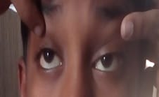
Myasthenia gravis (MG) is a rare autoimmune disorder affecting neuromuscular junction by muscle weakness. Myasthenia gravis can be generalized or localized as ocular myasthenia gravis. Case presentation: We report an 8-year-old boy who presented with 10 days history of drooping of both eyelids and 8 days history of diplopia. Examination revealed bilateral ptosis. A diagnosis of Juvenile Ocular Myasthenia gravis was made when symptoms improved with intramuscular Edrophonium administration. He was commenced on oral Neostigmine at a dose of 2mg/Kg/ day,4 hourly in divided doses and is on regular follow up and had a good response. Conclusion: Ocular Myasthenia gravis (OMG) is a rare disease in itself. A high index of suspicion is required in a juvenile as it is even rarer.

Keywords: Ocular myasthenia gravis, Juvenile myasthenia, Oral Neostigmine, Ptosis, Diplopia

Corresponding Author How to Cite this Article To Browse
Chaitra MC, Assistant Professor, Department of Ophthalmology, Sri Devaraj Urs Medical College, Kolar, Karnataka, India.
Email:
Usha BR, Nandhini K, Chaitra MC. Seeking spark- ocular myasthenia gravis in a juvenile – an Indian case report. Trop J Ophthalmol Otolaryngol. 2020;5(7):190-193.
Available From
https://opthalmology.medresearch.in/index.php/jooo/article/view/162

Manuscript Received Review Round 1 Review Round 2 Review Round 3 Accepted
2020-10-09 2020-10-14 2020-10-16 2020-10-19
Conflict of Interest Funding Ethical Approval Plagiarism X-checker Note
No Nil Yes 9%

© 2020 by Usha BR, Nandhini K, Chaitra MC and Published by Siddharth Health Research and Social Welfare Society. This is an Open Access article licensed under a Creative Commons Attribution 4.0 International License https://creativecommons.org/licenses/by/4.0/ unported [CC BY 4.0].

Introduction

Myasthenia gravis(MG) is an autoimmune disorder characterized by muscle weakness and fatigability [1,2,3]. It is due to autoantibodies causing receptor depletion at the neuromuscular junction, which results in impaired acetylcholine function [2,4]. It causes impaired nerve function and muscle weakness [1,3,5]. Myasthenia gravis can be generalized or localized as ocular myasthenia gravis [3,4,6].

Juvenile myasthenia gravis (JMG) is a rare condition and is distinct from adult-onset disease [4,7]. Ocular myasthenia is characterized by easy fatigability of extraocular muscles, commoner in females. Diagnosis is the Tensilon test or Neostigmine test, clinically [1,8]. Investigations include acetylcholine receptors autoantibodies titers. Treatment includes the use of anticholinesterase, immunosuppressants, plasmapheresis, and thymectomy when indicated [6,7,9,10].

Case Report

An 8-year-old boy presented with a history of drooping of upper lids of both eyes for 10 days. It was more in the left eye than the right eye. It was acute in onset and progressive. His mother had noticed that the drooping of upper lids was not constant and was not present as soon as the boy opened his eyes, after his sleep.

It worsened as the day progressed. He also started complaining of double vision for the past 8 days, which was variable and was not present in any particular gaze. He didn’t have any trauma before these symptoms developed. There was no associated fever or headache. His vision was good before and after his illness. He had no difficulty in breathing or swallowing or drooling of saliva. He did all his routine chores by himself and played well.

He didn’t feel tired or the week after exertion. There was no history of pain around the eyes or other facial muscles. There was no history of abnormal jerks or movements of the facial muscles. Pregnancy and neonatal histories were unremarkable. All aspects of motor development were normal. He was doing well academically. There was no similar illness in the family.

On examination, the boy was conscious cooperative, and well oriented to time, place, and person. His vitals were stable.

His systemic examination was normal. Ocular examination was done and his visual acuity was 6/6 N6 in both the eyes. Ocular alignment was orthophoric. Ptosis was present in both eyes (more in the left eye).

Ptosis measurements showed diurnal variation (worse in the evening). The fatigability test was positive in BE. Ptosis improved after the sleep test and ice pack test in both eyes (Figure 1). His pupils were 3mm round regular reacting equally to both direct and indirect light reflex. All uniocular and binocular extraocular movements were restricted in both the eyes and showed diurnal variation which worsened in the evening (Figure 2). Fundus was normal in both eyes.

opthal_162_01.jpg

Before the ice pack test.

opthal_162_02.jpg

After the ice pack test.

Fig-1: Condition before and after the Ice Pack test.

opthal_162_03.jpg


opthal_162_04.jpg

opthal_162_05.jpg opthal_162_06.jpg

Fig-2: Extraocular movements were restricted and showed a fluctuating pattern.

Intraocular pressure was done by applanation tonometry was 12mmHg in both the eyes. Clinical diagnosis of Ocular myasthenia gravis was made and an Edrophonium test was done and it showed significant improvement.

Complete blood count, Erythrocyte sedimentation rate, thyroid function test, blood sugar levels were normal. CT chest showed no thymoma. Thyroid function tests (T3, T4, and TSH) done were within the normal range.

Treatment: He was commenced on oral Neostigmine at a dose of 2mg/Kg/ day,4 hourly in divided doses and is on regular follow up and had a good response.

Discussion

Myasthenia gravis affects all ages and races [1,2,3]. Its incidence is 4-5 / 1,00,000. MG is more in young women and older men. Nearly 75% of MG initially present with ptosis, diplopia, or both. Nearly 60% of patients eventually develop generalized MG within 2 years.

JMG is rare and < 10-15% of all MG cases [4]. JMG is a 1-5 per million case per year. Over two-thirds of all MG begins with symptoms relating to their vision.

Pathophysiology: MG is characterized by the rapid fatigability of striated muscles [5,6]. The most common cause is an immune-mediated neuromuscular junction blockade [4,7]. The anti-acetylcholine receptor antibodies are the result of the T-cell dependent B-cell response.

Pathogenic anti-acetylcholine receptor antibodies are polyclonal and heterogeneous complicating the treatment using specific immunotherapy [2]. The role of the thymus and T lymphocyte in the production of antibodies is complex. Many patients improve after thymectomy

Acetylcholine is normally released into the synaptic cleft at the postsynaptic motor endplate is less responsive than normal. There is a reduced number of acetylcholine receptors due to antibodies. The pathophysiology of ocular myasthenia is similar to generalized myasthenia.

Why ocular muscles are frequently involved in MG is unclear. The reason may be due to morphologic and physiologic differences from limb muscles. Extraocular muscles are single innervated twitch fibers and have high firing frequencies.

In the pediatric population, there are 3 variants of MG.

  1. Transient neonatal
  2. Congenital
  3. Juvenile 4.

Transient neonatal is due to the passive transmission of autoimmune antibodies from a mother with myasthenia. Congenital Myasthenia is a rare disease due to the inheritance of defective neuromuscular junction synapse. Juvenile MG is the most common type of pediatric MG and subtype of MG. Juvenile MG has 2 variants.

  1. Generalised and
  2. Ocular.

It can occur at any age. It differs from adult-onset except that it frequently improves with age. The most common presentation is ptosis next common is diplopia in MG. these symptoms worsens as the day progresses. Other symptoms of generalized MG are slurred speech, dysphagia, feeling week, dyspnea, dysarthria.

Diagnostic tests are needed to confirm clinical suspicion include anti-AChR antibodies, electromyography (EMG) repetitive nerve stimulation (RNS) test, edrophonium tests [6]. Associated conditions including thymoma and thymic hyperplasia should be ruled out with appropriate investigations such as chest CT scan

Both ocular and generalized myasthenia gravis patients tend to have remissions and exacerbations at regular intervals. Treatment includes both medical and surgical6,8. Medications may include cholinesterase inhibitors such as Mestinon, steroids such as Prednisone, or other immunosuppressants used alone or in combination. Other modalities of treatment include plasmapheresis or IVIG (intravenous immunoglobulin) therapy which can be used either individually or in combination with other treatments [9].

These treatments offer only a temporary improvement and repeated treatments are necessary to sustain the effect. While thymectomy (removal of the thymus gland) is often recommended for patients with generalized MG, it is rarely used in purely ocular MG unless a thymoma is suspected [5,10].

Conclusion

Ocular myasthenia gravis is most likely to progress to generalized myasthenia, so regular follow up is required.

Ethics and consent

Written/ signed informed consent was obtained from the patient for the publication of this case and accompanying images. The patient’s confidentiality was also maintained throughout.

Reference

  1. Nair AG, Chhablani PP, Venkataramani DV, Gandhi RA. Ocular myasthenia gravis- A review. IJO. 2014;62(10)985-991.
  1. Lin CW, Chen T,Jou J, Woung L. Update on ocular myasthenia gravis in Taiwan. TJO. 2018;8(2)67-73.
  2. Cao Y, Bu B. Childhood-Onset Myasthenia Grav is, a Troublesome Clinical Entity with Heteroge neity in China. Arch Neurol Neurosci. 2019;2(3).
  3. Liew WKM, Kang PB. Update on juvenile myasthenia gravis. Curr Opin Pediatr. 2013;25(6)694-700.
  4. Solano A, Jimeno V, Montoya L, Espinosa N. Pediatric ocular myasthenia gravis- Case report and literature review. Pan Am J Ophthalmol. 2019;1(1)16.
  5. Pineles SL, Avery RA, Moss HE, Finkel R, Blinman T, Kaiser L, et al. Visual and systemic outcomes in pediatric ocular myasthenia gravis. Am J Ophthalmol. 2010;150(4)453-459.
  6. Ionita CM, Acsadi G. Management of juvenile myasthenia gravis. Pediatr Neurol. 2013;48(2)95-104.
  7. Howard JF Jr, Utsugisawa K, Benatar M, Murai H, Barohn RJ, Illa I, et al. Safety and efficacy of eculizumab in anti-acetylcholine receptor antibody-positive refractory generalised myasthenia gravis (REGAIN)- a phase 3, randomised, double-blind, placebo-controlled, multicentre study. Lancet Neurol. 2017;16(12)976-986.
  8. Cornblath, Wayne T. Treatment of Ocular Myasthenia Gravis. APJO. 2018;7(4)257-259.
  9. Kusner LL, Puwanant A, Kaminski HJ. Ocular myasthenia- diagnosis, treatment, and pathogenesis. Neurol. 2006;12(5)231-239.