E-ISSN:2456-6454
P-ISSN:2581-4907
RNI:MPENG/2017/74152

Research Article

Vitrectomy

Tropical Journal of Ophthalmology and Otolaryngology

2020 Volume 5 Number 6 July-August
Publisherwww.medresearch.in

Prognostic factors in Pars Plana vitrectomy for Proliferative Diabetic Retinopathy

Rukmangathan K.1*, P. N.2, Ravichandran R.3, Chandrasekaran R.4
DOI: https://doi.org/10.17511/jooo.2020.i06.01

1* Keerthivarman Rukmangathan, Associate Professor, Department of Ophthalmology, Annapoorana Medical College, Salem, Tamil Nadu, India.

2 Nandhakishore P., Consultant, Aravind Eye Hospital, Theni, Tamil Nadu, India.

3 Ramusiddharthan Ravichandran, Consultant, Sri Ramanamaharishi Eye Hospital, Thiruvannamalai, Tamil Nadu, India.

4 Ranjan Chandrasekaran, Consultant, Annai Eye Hospital, Ramanathapuram, Tamil Nadu, India.

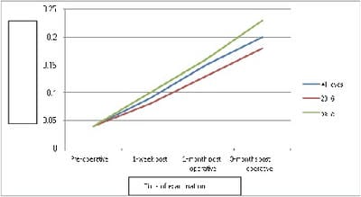
Objective: To determine the factors associated with good visual outcome in eyes undergoing pars plana vitrectomy (PPV) for proliferative diabetic retinopathy (PDR). Furthermore, the objective of the study was to compare the clinical outcome and complications of standard 20 gauge vitrectomy with 23-gauge transconjunctival sutureless vitrectomy. Materials and Methods: This was a prospective interventional study performed on patients presenting at the Retina clinic of a tertiary eye care hospital in Salem, Tamil Nadu between October 2018 to November 2019. All eyes undergoing PPV for complications of PDR and having adequate follow were included. The patients were divided into two groups, one group undergoing standard 20 gauge vitrectomy and the other group undergoing 23 gauge transconjunctival sutureless vitrectomy. Results: A total of Forty-nine eyes (25 eyes underwent standard 20 gauge vitrectomy and 24 eyes underwent 23 gauge transconjunctival sutureless vitrectomy). Visual acuity improved significantly following PPV (p<0.0001) and this improvement was seen in both 20 gauge group (p=0.0004) and in 23 gauge group (p=0.00005). There was no significant difference in best-corrected visual acuity noted between the two groups. However, eyes that underwent 23 gauge vitrectomy tended to gain vision earlier when compared to eyes that underwent 20 gauge vitrectomy. Complications following vitrectomy did not differ significantly between 20 gauge and 23 gauge group. Conclusion: Visual acuity improved significantly following PPV across all indications. Visual acuity in eyes that underwent 23 gauge TSV tended to gain vision earlier than eyes that underwent standard 20 gauge vitrectomy.

Keywords: Proliferative diabetic retinopathy, Pars plana vitrectomy, Non-proliferative diabetic retinopathy

Corresponding Author How to Cite this Article To Browse
Keerthivarman Rukmangathan, Associate Professor, Department of Ophthalmology, Annapoorana Medical College, Salem, Tamil Nadu, India.
Email:
Rukmangathan K, Nandhakishore P, Ramusiddharthan R, Chandrasekaran R. Prognostic factors in Pars Plana vitrectomy for Proliferative Diabetic Retinopathy. Trop J Ophthalmol Otolaryngol. 2020;5(6):116-124.
Available From
https://opthalmology.medresearch.in/index.php/jooo/article/view/148

Manuscript Received Review Round 1 Review Round 2 Review Round 3 Accepted
2020-07-16 2020-07-31 2020-08-13 2020-08-26
Conflict of Interest Funding Ethical Approval Plagiarism X-checker Note
No Nil Yes 7%

© 2020 by Keerthivarman Rukmangathan, Nandhakishore P., Ramusiddharthan Ravichandran, Ranjan Chandrasekaran and Published by Siddharth Health Research and Social Welfare Society. This is an Open Access article licensed under a Creative Commons Attribution 4.0 International License https://creativecommons.org/licenses/by/4.0/ unported [CC BY 4.0].

Introduction

India has currently the world’s largest diabetic population and the majority of them are unaware of their diabetic status [1]. Those who are aware do not realize the importance of consistent follow-up with physicians and most of the physicians are not trained to detect and refer sight-threatening diabetic retinopathy (DR). The DR is a complication of diabetes which causes damage to blood vessels of the retina. It is one of the leading causes of newly diagnosed legal blindness amongst working people. DR is broadly classified into non-proliferative diabetic retinopathy (NPDR) and proliferative diabetic retinopathy (PDR).

Type 2 DM is commonly associated with diabetic retinopathy with macular edema [2]. Diabetic macular edema is the most frequent cause of visual impairment in diabetic patients which can occur at any stage of DR, but it is more common in advanced stages (70% of eyes with PDR) [3]. It is characterized by foveal edema, exudates, or ischemia. Diffuse retinal edema is due to extensive capillary leakage, and localized edema due to focal leakage from microaneurysms and dilated capillary segments [4]. These data are alarming because most diabetics develop some retinopathy in the course of their lifetime.

The development and progression of diabetic retinopathy can be prevented by strict glycaemic control and other associated risk factors, such as hypertension and renal disease [5-7] which accelerates the disease process. Initial stages of diabetic retinopathy require treatment like laser photocoagulation (focal, grid, or pan-retinal laser photocoagulation) or the use of intravitreal anti-VEGF or corticosteroids for CSME and proliferative changes to restore vision. surgical intervention like PPV is indicated for advanced PDR.

PPV in diabetic retinopathy can be associated with a significant risk of complications. Intraoperative complications of vitrectomy include peripheral retinal dialysis, peripheral tears, vitreous or retinal incarceration in the wound, corneal erosions, filamentary keratitis, bullous keratopathy, damage to the lens by the direct touch of instruments, and the solutions used; intraoperative hemorrhage, retinal tears, retinal detachment, and posterior breaks can also occur [8,9]. Post-operative complications after PPV include residual hemorrhage from surgery, retained blood within vitreous, continued hemorrhage from bleeding sites, retinal

traction, bullous retinal detachment, anterior hyaloid proliferation, a progression of cataract, corneal decompensation, hypotony, post-operative glaucoma, and endophthalmitis [10-11].

Although sophisticated instruments and lasers have been developed for vitreoretinal surgery still the patient needs to undergo 20 gauge sclerotomies and postoperative morbidity because of sutured wounds. Sutureless self-sealing sclerotomies were initially proposed by Chen [12] has gained in popularity.

Over the years modifications in vitrectomy instruments such as 23-gauge transconjunctival sutureless vitrectomy (TVS) introduced by Eckardt [13] in 2005 which had less instrument flexibility allowing greater ocular rotation and the ability to perform a more complete peripheral vitrectomy.

In this present investigation, an attempt has been made to document the prognostic factors in PPV in patients presenting with diabetic retinopathy (DR) at a tertiary eye care facility in Tamil Nadu, India. Also, the surgical outcomes and complications of standard 20 gauge vitrectomy are compared with 23-gauge transconjunctival sutureless vitrectomy.

Materials and Methods

Study design and sample size: A prospective interventional study was conducted in the Department of Ophthalmology, Annapoorana Medical College and Hospital, from the period of March 2019 to April 2020, Salem, Tamil Nadu. This study was approved by the institutional Ethics Committee. Over a period of 13 months, 72 patients underwent pars plana vitrectomy for complications of diabetic retinopathy at Annapoorana Medical College and Hospital and were considered for inclusion in the current study. Of these, 23 patients were excluded due to one or more exclusion criteria, hence 49 patients (49 eyes) were included in the study.

Inclusion criteria

  1. Presented with proliferative diabetic retinopathy and undergoing 23G pars plana vitrectomy or standard 20 gauge vitrectomy to manage complications of proliferative diabetic retinopathy.
  2. A patient who completed 3 months of follow-up.

Exclusion criteria

  1. Did not provide informed consent.

  1. Presented with other retinal vascular disorders, epiretinal membrane, or macular hole not attributable to diabetes.
  2. Patients with pre-existing conditions such as age-related macular degeneration and advanced glaucoma which adversely affect visual outcomes.
  3. Any medical condition for which PPV was contraindicated.
  4. Data of patients who had not completed a three-month follow-up were excluded from the analysis.

The patients were divided into two groups, one group undergoing standard 20 gauge vitrectomy and the second group undergoing 23 gauge transconjunctival sutureless vitrectomy (TSV).

A complete medical and ocular history was taken at the baseline visit, including duration of diabetes, treatment history, control of diabetes, and other co-existent morbidities.

All the patients underwent a detailed ophthalmic examination and All patients received an explanation of the procedure and it’s possible complications and written informed consent was obtained. The main outcome measure evaluated was visual acuity. Good visual acuity was defined as best-corrected visual acuity of 6/24 or more with clear media and well-attached retina. The secondary outcome evaluated was an anatomical success following vitrectomy which was defined as improvement in BCVA compared to preoperative visual acuity.

Also, clinical outcomes and complications were compared between standard 20Gvitrectomyversus 23G transconjunctival sutureless vitrectomy (TSV). All obtained data were analyzed by using an unpaired student t-test for intergroup analysis of mean and the Chi-square test was used for proportions. Differences between values were considered to be statistically significant if P-value is ≤ 0.05.

Results

A total of 49 patients enrolled in the study, 38 (78%) patients were male and 11 (22%) patients were female (Figure 1). The mean age of the patients was 53.06 ± 7.5 years (range 36 to 71 years) and Pars plana Vitrectomy was performed for the right eye in 29 instances (59%) and for the left eye in 20 instances (41%). (Figure 1).

opthal_148_01.jpg

Fig-1(A): Gender distribution.

opthal_148_02.jpg

Fig-1(B): Age characterization.

opthal_148_03.jpg

Fig-1(C): Laterality.

Fig-1: Gender distribution, Age characteristics, and Laterality of the eyes undergoing pars plana vitrectomy for proliferative diabetic retinopathy.

In this current study, 37 patients (75%) had suffered from diabetes mellitus for a period of one to 10 years, 10 patients (21%) had diabetes ranging from 11 to 20 years and two patients (4%) had diabetes for a period of 30 to 40 years (Figure 2).

opthal_148_04.jpg

Diabetes Mellitus -DM, Hypertension –HT

Chronic kidney diseases – CKD, ischaemic heart disease -IHD

Fig-2: Duration of diabetes mellitus Coexisting


morbidity in patients undergoing pars plana vitrectomy for proliferative diabetic retinopathy.

A Total 41% of patients had presented with diabetic retinopathy, and the remaining 59% also had co-morbid conditions (34% - with hypertension and diabetes mellitus,4%-chronic kidney disease and diabetes mellitus, and 2%- ischemic heart disease and diabetes mellitus)(Figure 2) and 91% were on oral hypoglycemic agents (such as sulfonylurea’s and metformin) and 9% were on insulin. A total of 25 eyes (51%) underwent 20G pars plana vitrectomy and 24 eyes (49%) underwent 23G sutureless pars plana vitrectomy and 28 eyes (57%) underwent at least one sitting of laser photocoagulation prior to pars plana vitrectomy whereas 21 eyes (43%) were not given any prior laser. Among the 40 eyes (82%) were given intravitreal bevacizumab (Avastin™) before pars plana vitrectomy whereas nine eyes (18%) were not given any intravitreal injections. Indication and type of surgery performed for 49 study subjects were included (Table 1).

Table-1: Indication for Pars Plana Vitrectomy and the type of surgery performed in patients with proliferative diabetic retinopathy.

Indications Number of eyes (% of total) Type of surgery performed No of patients
VH 31 (63%) PPV + MP + EL + SOI 29
TRD 6 (12%) PPV + MP + EL + GAS 3
VH+TRD 9 (18%) PPV + EL 4
VH+TRD+RRD 1 (2%) PPV + MP + EL + SOI + IOL 9
VH+ MACULAR EDEMA 1 (2%) PPV + MP + EL + GAS +IOL 3
TRD+RRD 1 (2%) PPV + EL + IOL 1

VH – Vitreous haemorrhage
TRD – Tractional retinal detachment
RRD – Rhegmatogenous retinal detachment
PPV- Pars Plana Vitrectomy
MP- Membrane Peeling
SOI-Silicon oil
IOL – Intraocular lens

Comparison of pre-operative and post-operative visual acuity

Comparison of 20 G and 23 G pre and post-operative visual acuity: In the 20 G-group the mean preoperative visual acuity (in decimals) was 0.04± 0.03(~2/60) while the mean postoperative

visual acuity(in decimals) at 1 week was 0.08 ± 0.06 (~5/60), which difference was statistically significant (‘t’ [df : 48] = 2.9 ; p = 0.004). At the end of the 1-month postoperative period, the mean visual acuity (in decimals) was 0.13 ±0.12 (~6/60) which value was statistically significantly better than the mean preoperative value (‘t’ [df:48] = 3.63; p= 0.007). At the 3 months post-operative review, the mean visual acuity (in decimals) was 0.18 ± 0.17 (~6/36) which was statistically significantly better than mean preoperative visual acuity (‘t’ [df: 48]= 4.05; p= 0.0002).

In the 23G group, the mean preoperative visual acuity (in decimals) was 0.04±0.03 (~2/60) while the mean visual acuity at 1 week post-surgery was 0.10 ± 0.08, (~ 6/60) which difference was statistically significant (‘t’ [df:46]= 3.1; p = 0.0029).

At the one month post-operative review the mean visual acuity (in decimals) was 0.16±0.10 (~ 6/36) which value was statistically significantly better than the mean pre-operative visual acuity (‘t’ [df:46]= 3.51; p= 0.0010). At the 3 months post-operative period, the mean visual acuity (in decimals) was 0.23 ± 0.20 (~ 6 /24) which was found to be statistically significantly better than the mean preoperative visual acuity (‘t’ [df: 46] = 3.72; p= 0.0005) (Table 2).

Table-2: Intra and Post-operative Complication following pars plana vitrectomy using different gauge sizes in patients with proliferative diabetic retinopathy.

  Indications Indications
VH TRD VH+ TRD Total VH TRD VH+ TRD Total
Intra Operative Bleeding - 2 1 3 - 1 - 1
Retinal Break 1 2 - 3 - 1 1 1
  Indications Indications
VH TRD VH+TRD Total VH TRD VH+TRD Total
Recurrent VH 1 1 - 2 - - - -
Retinal Break 1 - - 1 - 1 - 1
Macular edema - - 1 1 - - 1 1
Combined RRD+TRD - - - - - 1 - 1

In this study, the improvement in visual acuity after surgery was compared between 25 eyes (51%) that underwent 20 gauge pars plana vitrectomy versus 24 eyes (49%) that underwent 23 gauge pars plana vitrectomy. The mean post-operative best-corrected visual acuity (BCVA) (decimals) at 3 months in the 20 gauge group was 0.18± 0.17 (~6/36) while the mean BCVA (decimals) in the 23 gauge group was 0.23 ± 0.22 (~6/24).


When the mean visual acuity (in decimals) in 20 gauge group was compared with that in the 23 gauge group 1 week after surgery the difference was not statistically significant (p = 0.4). Similarly no significant differences were noted when compared at the 1 month (p = 0.3) and 3 month (p = 0.4) post-operative review (Figure 3).

Visual acuity in decimals

opthal_148_05.jpg

Fig-3: Mean pre-operative and post-operative visual acuity in patients with proliferative diabetic retinopathy undergoing pars plana vitrectomy.

In addition to the comparison of 20 G and 23 G post-operative visual acuity, the study carried over the application of silicon oil without silicon oil. The mean visual acuity (in decimals) in the silicone oil group was compared with that in the group without silicone oil at 1-week post-operative review, the difference was statistically significant (‘t’ [df:47]= 2.4692; p= 0.0172), but the difference was not statistically significant at the 1 month (p= 0.12) and 3 months (p = 0.006) post-operative review.

In this study furthermore, the comparison between the post-operative visual acuity with pre- operative application of laser and bevacizumab. The mean BCVA in the laser group and bevacizumab was compared with that in the group without laser and bevacizumab at the 1-week post-operative review the difference was not statistically significant (p =0.6) and (p=0.7) respectively. Similarly, no significant differences were noted when compared with the BCVA at the one-month (p= 0.8), (p = 0.3) and 3-months (p=0.4) , (p= 0.5) review times respectively.

Comparison of pre and post-operative complication:In this study, of the 49 eyes which underwent pars plana vitrectomy, four eyes (8%) developed intraoperative bleeding (three eyes from the 20G group and one eye from the 23 G group) and four eyes (8%) developed iatrogenic retinal

break (three eyes from the 20G group and one eye from the 23G group). On Post-operative complications two eyes (from 20G group) developed recurrent vitreous hemorrhage, two eyes had retinal break detected post-operatively (one each from 20G and 23G group), two eyes (one each from 20G and 23G group) presented with severe macular edema, and one eye (from 23 G group) developed combined rhegmatogenous and tractional retinal detachment (Table 2).

In this study, 6 eyes from 20 gauge group and 2 eyes from 23 gauge group developed complications intraoperatively, this difference was not statistically significant. In the post-operative visits, 4 eyes from 20 gauge group and 3 eyes from 23 gauge group had complications, this difference was not statistically significant.

Discussion

Pars plana vitrectomy (PPV) in proliferative diabetic retinopathy (PDR) can be effective in clearing media opacities (such as blood) and relieving traction from fibrovascular tissue. However, PPV in eyes with PDR can be associated with severe and vision-threatening complications, and the final outcome, in terms of anatomical and visual forms depends on several factors. Hence, it is important to determine the visual prognosis associated with PPV for various indications in proliferative diabetic retinopathy so that an optimum course of treatment can be chosen.

A landmark trial was conducted in the year 1976, known as the Diabetic Retinopathy Vitrectomy Study (DRVS) [14], in which two randomized trials were done to determine whether 1) Early vitrectomy was preferable to delayed vitrectomy in eyes with severe vitreous hemorrhage due to PDR and 2) Early vitrectomy is preferable in eyes with PDR without vitreous hemorrhage. The study concluded that early vitrectomy is recommended for eyes with severe vision loss from non-clearing vitreous hemorrhage of at least one-month duration in patients with type 1 DM, or in one-eyed patients regardless of the type of diabetes.

The current study the patients who underwent PPV for treatment of complications due to diabetic retinopathy were mostly male patients (78%) and the laterality of the eyes was almost equally matched undergoing PPV. The mean age of the patients in the study who underwent vitrectomy was 53 years, while 75% of the patients who enrolled in


the study suffered from diabetes for around 10 years. In this study, 41% of the patients had other associated (co-morbid) systemic diseases along with diabetes. Most of the patients who underwent vitrectomy had type II diabetes (90%). The present study was also on par with Ferreira et al [15].

Various studies related to indications for vitrectomy in diabetic retinopathy by Thompson et al [16], Tony et al [17], and Helbig et al [18], found that severe non-clearing vitreous hemorrhage, tractional retinal detachment (TRD), combined tractional and rhegmatogenous retinal detachment and fibrovascular proliferation as indications for vitrectomy due to complications of diabetic retinopathy, dense pre macular hemorrhage. In the current study, the most frequent indication for PPV was found to be non-clearing vitreous hemorrhage (63%) (which was similar to other studies), followed by combined tractional retinal detachment with vitreous hemorrhage (18%); 12% of eyes had TRD, 2% of eyes had recalcitrant macular edema and another 2 % had combined tractional and rhegmatogenous retinal detachment.

Coming to surgical procedures used, Qamar et al [19], performed PPV for patients with TRD without retinal break peeling with endolaser. These authors concluded that TRD without retinal breaks can be treated by PPV without internal tamponade. In another study, Castellarin et al [20] concluded that PPV with silicone oil infusion is useful in severely affected eyes with PDR even in the presence of rubeosis iridis and neovascular glaucoma, and also in cases of previously failed vitrectomy. In yet another study, Canan et al [21] observed that the combined operation of PPV, phacoemulsification, and intraocular lens implantation was safe and effective in patients with PDR; visual outcome and complications depended on underlying posterior segment pathology and were not related to the combined procedure technique. Interestingly, Avitabile et al [22] compared PPV with pan-retinal photocoagulation or severe PDR. In their study, one group of patients underwent membrane peeling with internal limiting membrane peeling while other groups underwent laser photocoagulation. These investigators concluded that surgery can be deferred in eyes with TRD not involving the macula until progression threatens the vascular center.

Modarres et al [23] evaluated the use of preoperative intravitreal bevacizumab in patients undergoing vitrectomy for proliferative diabetic retinopathy. In their study, 22 eyes were given

preoperative intravitreal bevacizumab and 18 eyes were not given any prior injections. These authors concluded that intravitreal bevacizumab before PPV decreased the rate of postoperative vitreous hemorrhage and improvement in visual acuity. Yeung et al [24] in their study concluded that preoperative intravitreal injection of bevacizumab was useful in reducing early postoperative vitreous hemorrhage in vitrectomy for diabetic tractional detachment. In the present study, in 40 eyes preoperative intravitreal bevacizumab was given and in nine eyes preoperative bevacizumab was not given. In the postoperative period, only one eye (2.5%) developed early vitreous hemorrhage in the bevacizumab group and two eyes (22%) developed vitreous hemorrhage in the group without bevacizumab.

As with any surgery, surgical complications may be encountered when performing PPV. Authors pointed that fibrovascular ingrowths at the sclerotomy sites West et al [25], and Dong et al [26] compared clinical outcomes and complications of 20-gauge vitrectomy versus 23-gauge transconjunctival sutureless vitrectomy in patients with PDR. In this study, intraoperatively, 8.6% of eyes developed a retinal tear in the 23-gauge group and 15.2 % of eyes in 20 gauge group, and during the post-operative period, early vitreous hemorrhage developed in 5.7% of eyes in 23 gauge group and 13.6% of eyes from 20 gauge group. Recurrent tractional retinal detachment complicated with fibrovascular growth was seen in 3% of eyes from the 20-gauge group and none in the 23-gauge group, neovascular glaucoma developed in 2.9% eyes from 23 gauge and 7.6% eyes from the 20-gauge group.

Sharif et al [27] in their study compared retinal breaks observed during 23-gauge transconjunctival sutureless vitrectomy versus conventional 20-gauge vitrectomy for PDR. Both the groups included 85 eyes each and in conclusion, it was found that the 23-gauge group had lower rates of retinal break formation compared to that of the 20-gauge group. In the present study, 25 eyes underwent standard 20-gauge vitrectomy and 24 eyes underwent 23-gauge transconjunctival sutureless vitrectomy. During the intraoperative period, three eyes (12%) developed bleeding in the 20-gauge group, and one eye (4%) in the 23-gauge group, retinal breaks were noted in three eyes in the 20-gauge group and one eye in the 23-gauge group. In the postoperative period, early vitreous hemorrhage was noted in one


eye in a 20-gauge group and none in the 23-gauge group and a combined tractional with a rhegmatogenous retinal detachment was noted in one eye in23 the gauge group. In the current study, the difference in the rate of complications between 20 gauge versus 23-gauge vitrectomy was not statistically significant.

Surgical outcomes of PPV for diabetic retinopathy complications may be varied. Machemer et al [28] in their study on vitrectomy for diabetic retinopathy, found that visual improvement occurred in 59% of the eyes in which the retina was stable, while in 25% of patients there was retinal detachment, which resulted in visual improvement in only 46% of the eyes; the total success rate was 51%. The main causes for bad prognosis were rubeosis of the iris and posterior retinal detachment with 42% of all eyes exhibiting some degree of rubeosis of iris. Most of the preoperative eyes (71%) had rubeosis presented postoperatively also with rubeosis. In another study, Amar et al [19] observed successful retinal reattachment in 92% of the operated eyes; in those patients, the retina was stable and attached till the end of one-year follow-up improvement in best-corrected visual acuity was seen in 75%; mean improvement in best-corrected visual acuity was 2.00+1.24 lines at baseline to 1.24+1.22 (p <0.05) at the end of follow up. Dong et al [26] in their study compared clinical outcomes between 23 trans-conjunctival sutureless vitrectomy (TSV) with standard 20 gauge vitrectomy where they compared best-corrected visual acuity between the two groups.

Limitation

  1. The follow-up period should have been longer, as the follow-up period in this study was only 3 months.
  2. PPV performed by a single surgeon would have been better, in this study it was performed by multiple surgeons.
  3. Indication for PPV was mostly vitreous hemorrhage and tractional retinal detachment, data of other indications for PPV for proliferative diabetic retinopathy would be more beneficial.

Conclusion

The authors concluded that there was no difference in best-corrected visual acuity between the two groups at each follow-up visit and concluded that 23 gauge (TSV) is as effective for proliferative diabetic

retinopathy as standard 20-gauge vitrectomy. In the present study, 90% of the patients were found to have stable, well-attached retina postoperatively, while 6% of the patients had a retinal detachment and 4% had persistent macular edema at the end of the follow-up period. These results were comparable to those of other studies, suggesting that PPV is a useful and successful surgical technique in PDR. Moreover, visual acuity improved significantly after surgery (p < 0.0001) and this improvement was seen in both 20-gauge group (p = 0.0004) and in 23-gauge group (p = 0.0005). There was no significant difference in best-corrected visual acuity noted between the two groups, however, eyes that underwent 23-gauge vitrectomy tended to gain vision earlier when compared to eyes that underwent 20-gauge vitrectomy).

What does the study add to the existing knowledge

Preoperative intravitreal injection of bevacizumab is useful in reducing early post-operative vitreous hemorrhage following PPV. 23-gauge TSV is as effective in treatment for proliferative diabetic retinopathy as standard 20-gauge vitrectomy. Visual acuity following gas tamponade should be assessed since most of the eyes underwent PPV with silicone oil. Eyes that underwent 23-gauge vitrectomy tended to gain vision earlier when compared to eyes that underwent 20-gauge vitrectomy.

Author’s contribution

Dr. Keerthivarman Rukmangathan: Concept, study design

Dr. P. Nandhakishore: Data analysis

Dr. Ramusiddharthan Ravichandran: Data analysis and Manuscript preparation

Dr. Ranjan Chandrasekaran: Manuscript preparation

Reference

  1. Diabetes Atlas. International diabetes foundation, Brussels, Belgium. 2nd ed, 2003 Chapter 7,7; South-east Asia. pp 255-259.
  2. Aiello L. Perspectives on diabetic retinopathy. Am J Ophthalmol. 2003;136(1)122-135.

  1. Klein R, Klein B, Moss S, Davis M, DeMets D. The Wisconsin Epidemiologic Study of Diabetic Retinopathy. Ophthalmol. 1984;91(12)1464-1474.
  2. Bresnick G. Diabetic Macular Edema. Ophthalmol. 1986;93(7)989-997.
  3. Diabetes Control and Complications Trial Research Groupa. Progression of Retinopathy with Intensive versus Conventional Treatment in the Diabetes Control and Complications Trial. Ophthalmol. 1995;102(4)647-661.
  4. UK Prospective Diabetes Study (UKPDS) Group. Intensive blood-glucose control with sulphonylureas or insulin compared with conventional treatment and risk of complications in patients with type 2 diabetes (UKPDS 33). The Lancet. 1998;352(9131)837-853.
  5. UK Prospective Diabetes Study Group. Tight blood pressure control and risk of macrovascular and microvascular complications in type 2 diabetes- UKPDS 38. BMJ. 1998;317(7160)703-713.
  6. Smiddy W, Flynn H. Vitrectomy in the Management of Diabetic Retinopathy. Surv Ophthalmol. 1999;43(6)491-507.
  7. Oyakawa R, Schachat A, Michels R, Rice T. Complications of Vitreous Surgery for Diabetic Retinopathy. Ophthalmol. 1983;90(5)517-521.
  8. Blankenship G, Cortez R, Machemer R. The lens and pars plana vitrectomy for diabetic retinopathy complications. Arch Ophthalmol. 1979;97(7)1263-1267.
  9. Eckardt C. Transconjunctival sutureless 23-gauge vitrectomy. Retina. 2005;25(2)208-211.
  1. Honavar S, Goyal M, Majji A, Sen P, Naduvilath T, Dandona L. Glaucoma after pars plana vitrectomy and silicone oil injection for complicated retinal detachments. Ophthalmol. 1999;106(1)169-177.
  2. Chen J. Sutureless Pars Plana Vitrectomy Through Self-sealing Sclerotomies. Arch Ophthalmol. 1996;114(10)1273.
  3. Arch Ophthalmol. Early Vitrectomy for Severe Vitreous Hemorrhage in Diabetic Retinopathy. Arch Ophthalmol. 1985;103(11)1644.
  4. Ferreira N, Pessoa B, Macedo M, Queirós P, Meireles A. Vitrectomy in diabetic retinopathy. European Vitreoretinal Society. 14, 2011.
  5. Thompson J, De Bustros S, Michels R, Rice T, Glaser B. Results of Vitrectomy for Proliferative Diabetic Retinopathy. Ophthalmol. 1986;93(12)1571-1574.
  6. Ho T, Smiddy W, Flynn H. Vitrectomy in the management of diabetic eye disease. Surv Ophthalmol. 1992;37(3)190-202.
  7. Helbig H. Surgery for Diabetic Retinopathy. Ophthalmologica. 2007;221(2)103-111.
  8. Qamar RMR, Saleem MI, Saleem MF. The outcomes of pars plana vitrectomy without endotamponade for tractional retinal detachment secondary toproliferative diabetic retinopathy. Int J Ophthalmol. 2013;6(5)671-674.
  9. Avitabile T, Bonfiglio V, Castiglione F, Castaing M, Contarino F, Mistretta A. Severe proliferative diabetic retinopathy treated with vitrectomy or panretinal photocoagulation. Can J Ophthalmol. 2011;46(4)345-351.

  1. Castellarin A, Grigorian R, Bhagat N, Del Priore L, Zarbin MA. Vitrectomy with silicone oilinfusion in severe diabetic retinopathy. Br J Ophthalmol. 2003;87(3)318-321.
  2. West JF, Gregor ZJ. Fibrovascular ingrowth and recurrent hemorrhage following diabetic vitrectomy. Br J Ophthalmol. 2000;84;822-825.
  3. Modarres M, Nazari H, Ghasemi Falavarjani K, Naseripour M, Hashemi M, Mehdi Parvaresh M. Intravitreal injection ofbevacizumab before vitrectomy for proliferative diabetic retinopathy. Eur J Ophthalmol. 2009;19(5)848-852.
  4. Yeung L, Liu L, Wu WC, Kuo YH, Chao AN, Chen KJ, et al. Reducing the incidence of early postoperative vitreous hemorrhage by preoperative intravitreal bevacizumab in vitrectomy for diabetic tractional retinal detachment. Acta Ophthalmol. 2010;88(6)635-640.
  1. Canan H, Sizmaz S, Altan YR. Surgical results of combined pars planavitrectomy and phacoemulsification for vitreous hemorrhage in PDR. Clin Ophthalmol. 2013;7;1597-1601.
  2. Park DH, Shin JP, Kim SY. Comparison of clinical outcomes between 23-gaugeand 20-gauge vitrectomy in patients with proliferative diabetic retinopathy. Retina. 2010;30(10)1662-1670.
  3. Issa SA, Connor A, Habib M, Steel DH. Comparison of retinal breaks observed during 23-gauge transconjunctival vitrectomy versus conventional 20-gauge surgery for proliferative diabetic retinopathy. Clin Ophthalmol. 2011;5;109-114.
  4. Machemer R, Blankenship G. Vitrectomy for proliferative diabetic retinopathyassociated with vitreous hemorrhage. Ophthalmol. 1981;88(7)643-646.