E-ISSN:2456-6454
P-ISSN:2581-4907
RNI:MPENG/2017/74152

Research Article

Gland

Tropical Journal of Ophthalmology and Otolaryngology

2020 Volume 5 Number 4 April
Publisherwww.medresearch.in

Schwannoma of the submandibular gland: A rare case report

Ravinder Raja K.1*, Basha Shaik F.2
DOI: https://doi.org/10.17511/jooo.2020.i04.03

1* Khetawat Ravinder Raja, Assistant Professor, ENT Department, Mahavir Institute of Medical Sciences, Vikarabad, Telangana, India.

2 Feroz Basha Shaik, Assistant Professor, ENT Department, Mahavir Institute of Medical Sciences, Vikarabad, Telangana, India.

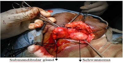
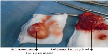
Schwannoma is a slow-growing, benign, encapsulated tumour of Schwann cells, which form myelin in peripheral nerve. It is the most common tumour of peripheral nerve, also known as neurilemma or neurinoma. Schwannomas of head and neck area are common and may arise from peripheral, central or autonomic nerve but salivary gland schwannomas are rare extracranial forms. The present study reports a rare case of schwannoma of the submandibular gland in a 42-year-old male patient which is presented with a painless mass in the submandibular region, treated by total excision of the submandibular gland. There were postoperative nerve deficit and no recurrence within 3months of follow up.

Keywords: Neurilemoma/Neurimoma, Tumour, Schwannomas, Submandibular gland

Corresponding Author How to Cite this Article To Browse
Khetawat Ravinder Raja, Assistant Professor, ENT Department, Mahavir Institute of Medical Sciences, Vikarabad, Telangana, India.
Email:
Raja KR, Shaik FB. Schwannoma of the submandibular gland: A rare case report. Trop J Ophthalmol Otolaryngol. 2020;5(4):96-99.
Available From
https://opthalmology.medresearch.in/index.php/jooo/article/view/139

Manuscript Received Review Round 1 Review Round 2 Review Round 3 Accepted
2019-04-02 2019-04-12 2019-04-17 2019-04-22
Conflict of Interest Funding Ethical Approval Plagiarism X-checker Note
No Nil Yes 8%

© 2020 by Khetawat Ravinder Raja, Feroz Basha Shaik and Published by Siddharth Health Research and Social Welfare Society. This is an Open Access article licensed under a Creative Commons Attribution 4.0 International License https://creativecommons.org/licenses/by/4.0/ unported [CC BY 4.0].

Introduction

Schwannomas are benign, solitary and well-differentiated tumours originating from Schwann cells [1]. Nearly 45% of all schwannomas occur in the head and neck area, where they may originate from any of the peripheral, cranial or autonomic nerves [2].

These benign tumours occur regardless of age or sex and are painless, insidious and slow-growing [3]. So, they are of long duration at the time of the presentation and rarely show a rapid course [4].

The nerve of origin is not identified in around 10 to 40% of schwannomas [5]. Histopathological examination demonstrates two types of schwannomas cells: Antoni type A and Antoni type B. The Antoni type A are spindle-shaped cells with parallel rows of palisading nuclear organized in whorls and waves, while the Antoni type B consists of spindle cells haphazardly scattered in a delicate, fibrillar microcystic matrix. Most schwannomas contain a mixture of both Antoni type A and Antoni type B tissue [16,17].

Case Report

A 42 years old male presented to the department of otorhinolaryngology of Mahavir institute of medical sciences Vikarabad with painless swelling in right submandibular gland region forthe last 5 months and loss of sensation of taste since 2months on the right side. The swelling was insidious in onset and slowly progressive in nature and loss of sensation of taste. On physical examination of the neck, a single hard swelling of approximate size 3x2x1cm noted in the right submandibular region. The surface was smooth, well defined lower margin but poorly defined upper margin, firm in consistency, non-tender and slightly mobile. There was no palpable lymphadenopathy. There was no history of fever, sore throat or local trauma. Clinical diagnosis of chronic submandibular sialadenitis was made. Ultrasonography neck showed 26x28mm well defined heterogeneously hypoechoic solid lesion with minimal vascularity seen in the right submandibular gland -? Adenoma and advised FNAC. Fine needle aspiration cytology showed scanty cellularity comprising of small sheets of basaloid ductal cells with sharp borders, These cells are having a dark oval nucleus and scanty cytoplasm suggestive of chronic submandibular sialadenitis and advised excision biopsy for confirmation.

The patient was prepared for the surgery under general anaesthesia after taking consent for excision of the submandibular gland. On exploration of the submandibular region, a swelling was attached to the submandibular gland was found.

opthal_139_01.jpg

Fig-1: Swelling was observed in the submandibular gland.

After careful dissection and identification of adjacent anatomical structures total excision of submandibular gland along with swelling done.

opthal_139_02.jpg

Fig-2: Total excision of the submandibular gland.

Wound healing was normal and there was neurapraxia (class1) of Seddon’s classification.

Pathological report

  1. Revealed glandular structure with lobules of acini, ducts and blood vessels. Increased gland to stromal ratio, dilated congested capillaries and epithelial hyperplasia – submandibular sialadenitis.
  2. Revealed compact hypercellular (Antoni A) andmyeloid hypocellular (Antoni B) areas and verocay bodies with nuclear palisading of elongated cells having spindle-shaped, wamy nucleus. Blood vessels are thrombosed, hyalinised walls. Interspersed collagen filness seen – schwannoma.

These findings confirm the diagnosis and there was no recurrence within 3 months of follow up.


Discussion

Schwannoma, first described in 1908 by Verocay, commonly occurs between 30 and 50 years old [6]; however, our case was a 42-year-old man. Neurilemmoma/schwannoma is a benign tumour arising from and consisting solely of Schwann cells [6]. Approximately 25 to 45 percent of extracranial schwannomas present in the head and neck area; the most commonly affected regions are the temporal bone, lateral neck, and paranasal sinuses [7]. Among the cranial nerves, schwannomas can arise from the glossopharyngeal, accessory, and hypoglossal nerves, while the most common type is acoustic neurinoma differentiating from the vestibulocochlear nerve [7].

Schwannomas are usually solitary lesions; however, some are multiple lesions as part of neurofibromatosis type 1 [6].

Clinical evidence of the tumour usually does not present for a long time. The most common symptom is a slow-growing mass [1,4,7,8]. Neurological symptoms and pain are rare. Schwannomas can be seen at any age and women show dominance [8,9]. Malignant transformation is very rare [1,8-10]. Diagnostic investigations include computed tomography (CT), magnetic resonance imaging (MRI), ultrasound scan and FNA. MRI is the best choice in detecting the extent of the tumour and correlates well with the operative findings [11]. Biswas et al. have reported their 10 years of experience regarding extracranial head and neck schwannomas, and in their report, only 6% of patients could have been diagnosed preoperatively on the basis of clinical findings, CT and MRI scans, and FNA [12].

Schwannomas have specific MRI properties, including specific signs (split-fat sign, fascicular sign, target sign) and signal patterns (that is, isointense T1 signal relative to skeletal muscle; increased and slightly heterogeneous T2 signal) [13].

Diagnosis is confirmed by histopathology showing the presence of Antoni A and Antoni B cells, nuclear palisading, whirling of cells and Verocay bodies [7,12,13]. The present study believed this tumour originated in an autonomic nerve of the submandibular gland. The treatment of schwannomas is problematic. Because of resistance to radiotherapy, surgical excision is necessary for optimal treatment [8,9,14,15].

Kang et al. reported that more than half of the surgically treated cases exhibited postoperative neural deficits that were primarily caused by iatrogenic injury to either the nerve of the origin or adjacent neural ending [7]. In our case, there was neurapraxia (class1) of Seddon's classification.

The malignant potential of extracranial schwannomas and risk of recurrence after surgical resection is unclear [14], while in most studies investigating extracranial schwannomas, recurrence or malignant transformation of the tumour have not been reported [7,12].

At 3 months follow-up of the present case, there was no evidence of recurrence and prognosis was excellent. Nevertheless, while a malignant transformation of schwannoma is an exceptionally rare event, disregarding this possibility seems not to be a safe practice inlight of the fact that malignancy can occur even if rarely [7,10,12,14].

Conclusions

Schwannoma of the salivary gland is a particularly rare form of an extracranial neurogenic tumour. Histopathologic examination isinevitable because MR findings are not specific.

Schwannomas were said to have specific MRI properties, including specific signs (split-fat sign, fascicular sign, target sign). However, they are not always observed.Our findings indicate good prognosis in an unusual case of a submandibular gland schwannoma in a 42-year-old man treated by surgical excision with no recurrence within 3 months of follow-up.

Reference

01. Rootman J, Goldberg C, Robertson W. Primary orbital schwannomas. Br J Ophthalmol. 1982;66;194–204. doi: [Article][Crossref][PubMed][Google Scholar]

02. Das Gupta T, Brassfiel R, Strong E, Hajdu S. Benign solitary schwannoma (neurilemmoma). Cancer. 1969;24(2)355-366. doi: (196908) 24:2<355::AID-CNCR2820240218>3.0.CO;2-2 [Article][Crossref][PubMed][Google Scholar]

03. Diaz DD, Kennedy KS, Parker GS, White VJ. Schwannoma of the submandibular gland. Head Neck. 1991;13(3)239-242. doi: 1002/hed.2880130314 [Article][Crossref][PubMed][Google Scholar]


04. Dhupar A, Yadaw S, Dhupar V. Schwannoma of the hard plate- a rare case. Internet J Head Neck Surg. 2011;4(2). Available from: 4/2/11719 [Article][Crossref][PubMed][Google Scholar]

05. Gallo WJ, Moss M, Shapiro DN, Gaul JV. Neurilemmoma- review of the literature and report of five cases. J Oral Surg. 1997;35(3)235-236. [Crossref][PubMed][Google Scholar]

06. Marx RE, Stern D. In- Oral and maxillofacial pathology- a rationale for diagnosis and treatment. Waters LC, editor, Carol Stream- Quintessence Publishing Co Inc. 2003. Benign soft tissue tumors of mesenchymal origin, pp-395-461 [Crossref][PubMed][Google Scholar]

07. Kang GC, Soo KC, Lim DT. Extracranial non-vestibular head and neck schwannomas- a ten-year experience. Ann Acad Med. 2007;36(4)233-238. [Crossref][PubMed][Google Scholar]

08. Torossian JM, Beziat JL, AbouChebel N, Devouassoux-Shisheboran M, Fischer G. Extracranial cephalic schwannomas- a series of 15 patients. J Craniofac Surg. 1999;10;389-394. doi: 00001665-199909000-00003 [Article][Crossref][PubMed][Google Scholar]

09. Leu YS, Chang KC. Extracranial head and neck schwannomas- a review of 8-year experience. Acta Otolaryngol. 2002;122(4)435-437. doi: org/10.1080/00016480260000157 [Article][Crossref][PubMed][Google Scholar]

10. Piscioli F, Antolini M, Pusiol T, Dalri P, Lo Bello MD, Mair K. Malignant schwannoma of the submandibular gland- A case report. J Otorhinolaryngol Relat Spec. 1986;48 (3)156-161. doi: [Article][Crossref][PubMed][Google Scholar]

11. Karaca CT, Habesoglu TE, Naboglu B, Habesoglu M, Oysu C, Egeli E, Tosun I. Schwannoma of the tongue in a child. Am J Otolaryngol. 2010;31(1)46-48. doi: [Article][Crossref][PubMed][Google Scholar]

12. Biswas D, Marnane CN, Mal R, Baldwin D. Extracranial head and neck schwannomas- a 10-year review. Auris Nasus Larynx. 2007;34(3)353-359. doi: [Article][Crossref][PubMed][Google Scholar]

13. Beaman FD, Kransdorf MJ, Menke DM. Schwannoma- radiologic and pathologic correlation. Radiographics. 2004;24(5)1477-1481. doi: org/10.1148/rg.245045001 [Article][Crossref][PubMed][Google Scholar]

14. Langner E, Del Negro A, Akashi HK, Araujo PP, Tincani AJ, Martins AS. Schwannoma in the head and neck- retrospective analysis of 21 patients and review of the literature. Sao Paulo Med J. 2007;125(4)220-222. doi: [Article][Crossref][PubMed][Google Scholar]

15. Neville BM, Damm DD, Allen CM, Bouquot JE. In- Oral Maxillofacial Pathology 3, Neville BW, Damm DD, Allen CM, Bouquot JE, editor. St Louis- Elsevier. 2009, Soft tissue tumors; pp-507–570. [Crossref][PubMed][Google Scholar]

16. Amir R, Altman KW, Zaheer S. Neurilemmoma of the hard palate. J Oral Maxillofac Surg. 2002;60(9)1069-1071. doi: [Article][Crossref][PubMed][Google Scholar]

17. Yamazaki H, Kaneko A, Ota Y, Tsukinoki K. Schwannoma of the mental nerve- Usefulness of preoperative imaging- A case report. Oral Surg Oral Med Oral Pathol Oral Radiol Endod. 2004;97(1)122–126. doi: [Article][Crossref][PubMed][Google Scholar]