E-ISSN:2456-6454
P-ISSN:2581-4907
RNI:MPENG/2017/74152

Research Article

Disc Edema

Tropical Journal of Ophthalmology and Otolaryngology

2020 Volume 5 Number 2 February
Publisherwww.medresearch.in

Bilateral optic disc edema: causes and clinical features at a tertiary center in South India

Ijeri R.1*, Jyoti RC.2
DOI: https://doi.org/10.17511/jooo.2020.i02.03

1* Raghavendra Ijeri, Assistant Professor, Department of Ophthalmology, Sri B M Patil Medical College Hospital and Research Centre, Vijayapur, Karnataka, India.

2 Jyoti RC, Senior Resident, Department of Ophthalmology, Sri B M Patil Medical College Hospital and Research Centre, Vijayapur, Karnataka, India.

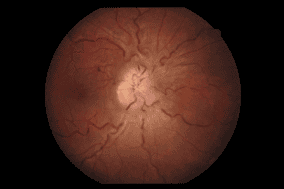
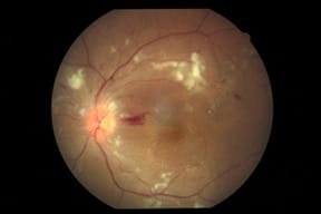
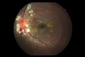
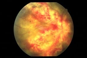
Aims: Bilateral optic disc edema: causes and clinical features at a tertiary center in South India. Bilateral Optic disc edema is one of the important fundus findings. Many times it indicates systemic pathology. The current study evaluated various causes of bilateral optic disc edema. Settings and Design: Prospective study. Methods and Material: This prospective study was conducted at a tertiary hospital in north Karnataka part of India (southern India). This study was conducted from June 2016 to November 2018. Detailed ocular examination including vision, slit-lamp examination, Visual field analysis, detailed fundus evaluation using an indirect ophthalmoscope, 78 Dioptre lens and fundus camera was done. Systemic investigations like blood examination and radiological investigations like Computerized tomography (C.T), Magnetic Resonance Imaging (MRI) were done whenever required. Results: A total of sixty-three cases with bilateral optic disc edema was recorded in this study. Thirty were females and thirty-three were males. Out of these, 36 cases had papilledema (increased intracranial pressure), 7 cases had optic neuritis, 6 cases of hypertensive retinopathy, 5 patients had Vogt Koyanagi Harada Syndrome (VKH), 3 had neuroretinitis, Cytomegalovirus (CMV) retinitis in 2 cases and in 4 cases causes could not be found. Total of 36 (57.14%) patients had papilledema, 7 (11.11%) had optic neuritis, 3 (4.8%) patient had neuroretinitis, 6 (9.52%) cases of hypertensive retinopathy, 5 (7.9%) patient had VKH, CMV retinitis in 2 (3.2%) and 4 (6.3%) idiopathic. Conclusions: Papilledema was the commonest cause followed by optic neuritis for bilateral optic disc edema.

Keywords: Bilateral Optic disc edema, Papilledema, Optic Neuritis, South India

Corresponding Author How to Cite this Article To Browse
Raghavendra Ijeri, Assistant Professor, Department of Ophthalmology, Sri B M Patil Medical College Hospital and Research Centre, Vijayapur, Karnataka, India.
Email:
Ijeri R, Jyoti RC. Bilateral optic disc edema: causes and clinical features at a tertiary center in South India. Trop J Ophthalmol Otolaryngol. 2020;5(2):55-59.
Available From
https://opthalmology.medresearch.in/index.php/jooo/article/view/124

Manuscript Received Review Round 1 Review Round 2 Review Round 3 Accepted
2020-01-06 2020-01-18 2020-01-22 2020-01-28
Conflict of Interest Funding Ethical Approval Plagiarism X-checker Note
No Nil Yes 4%

© 2020 by Raghavendra Ijeri, Jyoti RC and Published by Siddharth Health Research and Social Welfare Society. This is an Open Access article licensed under a Creative Commons Attribution 4.0 International License https://creativecommons.org/licenses/by/4.0/ unported [CC BY 4.0].

Introduction

Optic disc edema is an important ocular sign seen in many conditions. Some of the important causes associated with unilateral optic disc edema are optic neuritis (ON), anterior ischemic optic neuropathy (AION), compressive optic neuropathy, retinal vein occlusion, diabetic papillopathy. Cases with bilateral optic disc swelling are often associated with papilledema [increased intracranial pressure] (Figure 1) and hypertensive retinopathy [1] (Figure 2).

opthal_124_01.png

Fig-1: Papilledema.

opthal_124_02.jpg

Fig-2: Hypertensive retinopathy.

There are very few studies on the causes and clinical features of bilateral optic disc swelling in India and they had a very small sample size. Therefore, the purpose of this study was to determine the clinical manifestations and etiology of bilateral optic disc edema in Indian patients which would be helpful for clinicians to look for common causes of optic disc edema.

Optic disc edema occurs due to the slowing of axoplasmic transport. Axoplasmic stasis can be caused by ischemia, inflammation, certain toxins as well as compression.

The term papilledema is commonly referred to as a condition of the optic nerve due to raised intracranial pressure. The term optic disc edema can be used instead of papilledema, in case if the disc edema is not because of increased intracranial pressure.

Subjects and Methods

Setting: This study was conducted at the Department of Ophthalmology, in a tertiary hospital in north Karnataka part of India (southern India). Both inpatients and outpatients were included in the study.

Duration: This study was conducted from June 2016 to November 2018.

Type of study: This is an observational study.

Inclusion criteria: All consecutive cases of bilateral optic disc edema diagnosed at our hospital were enrolled for this study.

Exclusion criteria: Old cases of optic disc edema were not taken in this study.

Data collection and analysis: In the present study females were thirty in number and the numbers of males were thirty-three. Data were analyzed according to the percentage of the different diseases were calculated.

Ethical clearance: Ethical clearance certificate was obtained from the university ethical clearance committee.

Methodology: All the patients were subjected to a detailed history and ophthalmic examination included visual acuity, detailed slit lamp examination; fundus was examined using 78 diopter lens and indirect ophthalmoscope, color vision, fundus photography. Fundus fluorescein angiography (FFA) and visual fields were done whenever indicated. Computed Tomography (C.T.) and Magnetic resonance imaging (M.R.I) with or without contrast were done whenever required.

The diagnostic criteria were as follows: Optic Neuritis was diagnosed according to the criteria used in the Optic Neuritis Treatment Trial (ONTT) [2]. Papilledema was diagnosed when optic disc edema was present with ‘signs and symptoms, radiographic signs and lumbar puncture signs of raised intracranial pressure. Other diseases were diagnosed by their characteristic clinical and radiological features.


Results

The present study included sixty-three patients. Among sixty-three patients, thirty-three were males and thirty were females. The mean age of the patient was 42.50 (Range 15 to 70 years). The majority of the patients were between the ages group of 30 to 60 years.

In the present study most common cause for bilateral optic disc edema was Papilledema seen in 36 patients (57.14%); followed by optic neuritis 7 cases (11.11%), 6 cases of hypertensive retinopathy (9.52%), VKH 5 cases (7.9%), Neuroretinitis (Figure 3) seen in 3 cases (4.8%), CMV Retinitis (Figure 4) in 2 cases (3.2%) and in 4 cases cause could not be found (6.3%).

opthal_124_03.jpg

Fig-3: Neuroretinitis.

opthal_124_04.jpg

Fig-4 CMV Retinitis.

Causes for papilledema in the present study was seen in 36 cases: cerebral venous sinus thrombosis was seen in 14 cases (22.22%), meningitis in 11 (17.5%) cases, intracranial bleed in 9 (14.3%) cases and intracranial tumors in 2 (3.1%) cases. In the present study, only one patient with optic neuritis had Multiple sclerosis in the remaining six patients cause optic neuritis could not be found out.

Among meningitis out of 11 cases: 6 had tubercular meningitis, 4 had bacterial meningitis and one had viral meningitis. Among Intracranial tumors one was glioblastoma and the other was meningioma.

In patients with optic neuritis, VKH syndrome, neuro retinitis, and CMV retinitis vision loss were the primary symptom. All VKH Patients and three patients with optic neuritis had eye pain.

Discussion

Optic disc edema occurs due to the slowing of axoplasmic transport. Axoplasmic stasis can be caused by ischemia, inflammation, certain toxins as well as compression. The term papilledema is commonly referred to as a condition of the optic nerve due to raised intracranial pressure. The term optic disc edema can be used instead of papilledema, in case if the disc edema is not because of increased intracranial pressure [3].

The study tried to find out the most common cause and presentation of bilateral optic disc edema in our region. There are few studies regarding bilateral optic disc edema. The present study included only the new cases of bilateral optic disc edema and try to compare our results with the previously published literature.

Fundus picture of papilledema shows the blurring of optic disc margins and dilated, and tortuous veins as shown in figure1. In papilledema hemorrhages and exudates are seen within the one-disc diameter of the optic disc; hemorrhages and exudates more than one-disc diameter suggests other causes like uveitis and retinitis. In hypertensive retinopathy, optic disc edema is seen along with soft exudates, superficial hemorrhages, hard exudates at the macula and arteriovenous crossing changes as seen in Figure 2.

Figure 3 shows neuroretinitis in which soft exudates and hemorrhages are seen along with optic disc edema. Along with these findings is also associated with retinal thickening, hard exudates at the macula and subretinal fluid at the macula. In CMV retinitis areas of hemorrhage and necrosis are seen along blood vessels with disc edema as shown in figure 4.

In VKH patients with crowded discs, circulatory disturbances of the branches of the short posterior ciliary artery and/or centripetal branches from the peripapillary choroid due to severe choroidal inflammation may cause axonal flow stasis and secondary axonal swelling of the optic nerve [4-5].


One disease may have elevated intracranial pressure despite normal CT or an MRI finding is cerebral venous sinuous thrombosis. Thus, combining Magnetic resonance venography (MRV) with these standard imaging modalities would be useful when attempting to diagnose this condition. MRV can be used to confirm venous filling defects because it enables clear visu­alization of the major intracranial veins and cranial sinus without the use of a contrast agent.

Neuroretinitis is a particular form of optic neuropathy characterized by acute visual loss in the setting of optic disc swelling and hard exudate arranged in a star figure around the fovea. The primary process in neuro retinitis involves inflammation of optic disc vasculature causing exudation of fluid into the peripapillary retina [6].

Very early findings of neuro retinitis using spectral-domain (SD-OCT). SD-OCT imaging was performed on the first day of presentation for patients with presumed neuroretinitis. Observations included the presence of ‘peripapillary infiltrates’, thought to be collections of inflammatory cells in the vitreous abutting the optic nerve. These peripapillary infiltrates appear early in the course of neuro retinitis before the appearance of the macular star and are not seen in other entities associated with disc edema such as papilledema and anterior ischemic optic neuropathy. So, this may help in the diagnosis of neuro retinitis from other diseases [7].

Table 1: Demonstrates the major causes of optic disc edema in various studies.

Disease Japan [8] Japan [9] S. Korea [10] Present study
Papilledema 59% 48.5% 44.44% 66.7%
Optic Neuritis 2% 12.12% 22.22% 11.11%
Pseudo Papilledema 16% 18.18% 8.2% NIL
Uveitis 8% NIL NIL 7.9%(VKH)

Most of the recent studies about bilateral disc edema are from Asia. The recent data about this topic in western countries is very less because of the non-availability of studies on this topic. Western data available is older so comparison with this data will not be proper for analysis.

Papilledema was the most common cause for bilateral optic disc edema in the present study which was comparable to the previous studies. The common causes of papilledema were cerebral venous sinus thrombosis, meningitis. In other studies, the most common cause of papilledema was intracranial tumors.

This difference may be due to an increase in infective diseases in our country.

Previous studies [9-10] also had pseudo papilledema cases. The diseases like optic disc drusen, small optic disc, papillophlebitis were seen in these cases were not seen in the present study.

VKH was also reported by Iijima et [8] al in their paper it was also seen in the present study.

Neuroretinitis was peculiar to the present study which was not seen in other studies [8-10].

Conclusion

The current study would suggest getting an MRI scan done whenever there is only optic disc swelling. If it is associated with exudates at the macula, the subretinal fluid then fluorescein angiography and OCT should be performed.

Some of our patients were bedridden so visual acuity could not be recorded in such patients. So comparing vision in the different causes of optic disc edema was not possible. The disadvantages of the present study are the smaller sample size and study done at a single center. A multicentre study is required to know the actual statistics of optic disc edema. The positive aspect of this study was this was a prospective study whereas all the previous such studies were retrospective. This was a hospital-based observational study. The current study tried to find out the common causes of bilateral optic disc edema.

What does the study add to the existing knowledge?

This the first of its kind study from India. No other study had evaluated bilateral causes of optic disc edema in India. The present study more number of papilledema followed by optic neuritis.

Diseases like CMV retinitis and neuroretinitis were not reported in earlier studies which are seen in the present study. Optic disc drusen, small optic disc, papillophlebitis were not seen in the present study which was reported in the present study.

Author’s contribution

Both the authors Dr. Raghavendra Ijeri and Dr. Jyoti RC contributed equally in concepts, design, literature search, data acquisition, and manuscript preparation.


Reference

01. Van Stavern GP. Optic disc edema. Semin Neurol. 2007;27;233-243. doi: 10.1055/s-2007-979684 [Crossref][PubMed][Google Scholar]

02. Optic Neuritis Study Group. The clinical profile of optic neuritis. Experience of the Optic Neuritis Treatment Trial. Arch Ophthalmol. 1991;109(12)1673-1678. doi: 10.1001/archopht.1991.01080120057025 [Crossref][PubMed][Google Scholar]

03. Hayreh SS. Pathogenesis of optic disc edema in raised intracranial pressure. Prog Retin Eye Res. 2016;50;108-144. doi: 10.1016/j.preteyeres.2015.10.001 [Crossref][PubMed][Google Scholar]

04. Rajendram R, Evans M, Khurana RN, Tsai JH, Rao NA. Vogt- Koyanagi-Harada disease presenting as optic neuritis. Int Ophthalmol. 2007;27;217-220. doi: 10.1007/s10792-006-9026-5 [Crossref][PubMed][Google Scholar]

05. Nakao K, Abematsu N, Mizushima Y, Sakamoto T. Optic disc swelling in Vogt-Koyanagi-Harada disease. Invest Ophthalmol Vis Sci. 2012;53;1917-1922. doi: 10.1167/iovs.11-8984 [Crossref][PubMed][Google Scholar]

06. Purvin V, Sundaram S, Kawasaki A. Neuroretinitis- review of the literature and new observations. J Neuro Ophthalmol. 2011;31(1)58-68. doi: 10.1097/wno.0b013e31820cf78a [Crossref][PubMed][Google Scholar]

07. Zatreanu L, Sibony PA, Kupersmith MJ. Optical coherence tomography in neuroretinitis- epipapillary infiltrates and retinal folds. J Neuro Ophthalmol. 2017;37(2)176-178. doi: 10.1097/WNO.0000000000000501 [Crossref][PubMed][Google Scholar]

08. Iijima K, Shimizu K, Ichibe Y. A study of the causes of bilateral optic disc swelling in Japanese patients. Clin Ophthalmol. 2014;8;1269-1274. doi: 10.2147/OPTH.S61650 [Crossref][PubMed][Google Scholar]

09. Hata M, Miyamoto K. Causes and prognosis of unilateral and bilateral optic disc swelling. Neuro-Ophthalmol. 2017;41(4)187-191. doi: 10.1080%2F01658107.2017.1299766 [Crossref][PubMed][Google Scholar]

10. Jung JJ, Baek SH, Kim US. Analysis of the causes of optic-disc swelling. Korean J Ophthalmol. 2011;25(1)33-36. doi: 10.3341%2Fkjo.2011.25.1.33 [Crossref][PubMed][Google Scholar]